. 2020 Aug 6;5(15):e137869.
doi: 10.1172/jci.insight.137869.

Inhibition of DDR1 enhances in vivo chemosensitivity in KRAS-mutant lung adenocarcinoma

Affiliations

Inhibition of DDR1 enhances in vivo chemosensitivity in KRAS-mutant lung adenocarcinoma

Marie-Julie Nokin et al. JCI Insight. .

Abstract

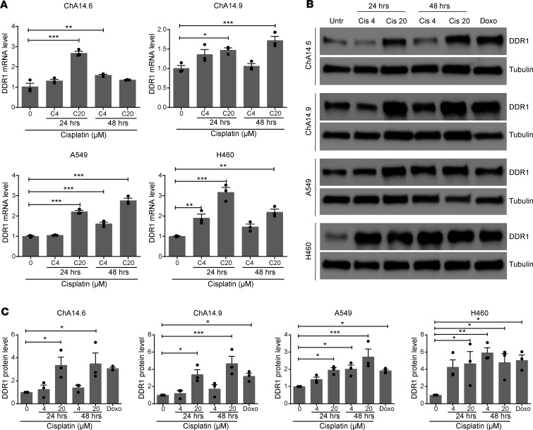
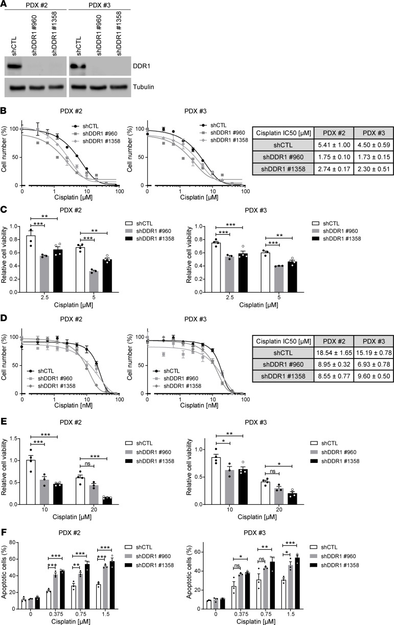
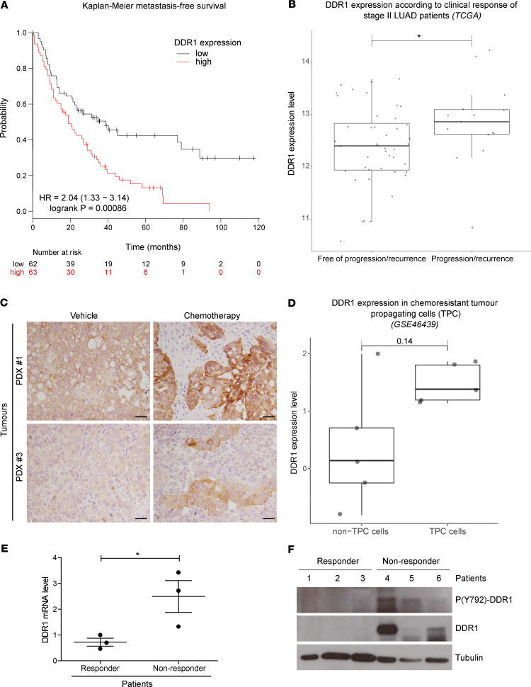
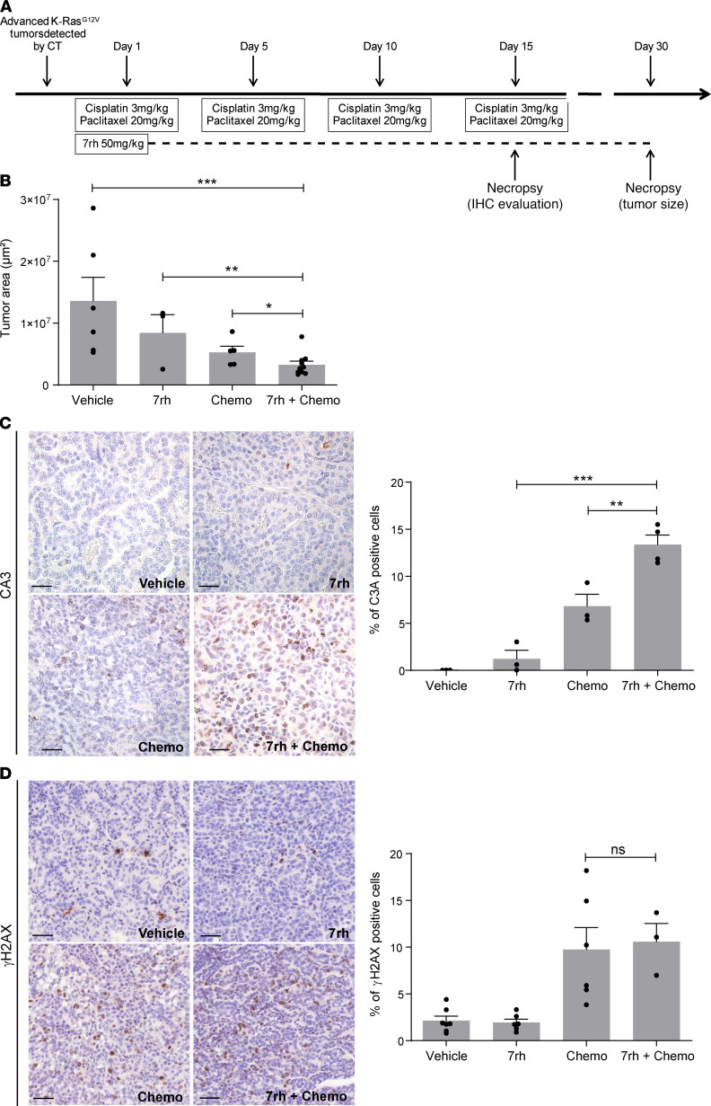
Platinum-based chemotherapy in combination with immune-checkpoint inhibitors is the current standard of care for patients with advanced lung adenocarcinoma (LUAD). However, tumor progression evolves in most cases. Therefore, predictive biomarkers are needed for better patient stratification and for the identification of new therapeutic strategies, including enhancing the efficacy of chemotoxic agents. Here, we hypothesized that discoidin domain receptor 1 (DDR1) may be both a predictive factor for chemoresistance in patients with LUAD and a potential target positively selected in resistant cells. By using biopsies from patients with LUAD, KRAS-mutant LUAD cell lines, and in vivo genetically engineered KRAS-driven mouse models, we evaluated the role of DDR1 in the context of chemotherapy treatment. We found that DDR1 is upregulated during chemotherapy both in vitro and in vivo. Moreover, analysis of a cohort of patients with LUAD suggested that high DDR1 levels in pretreatment biopsies correlated with poor response to chemotherapy. Additionally, we showed that combining DDR1 inhibition with chemotherapy prompted a synergistic therapeutic effect and enhanced cell death of KRAS-mutant tumors in vivo. Collectively, this study suggests a potential role for DDR1 as both a predictive and prognostic biomarker, potentially improving the chemotherapy response of patients with LUAD.

Keywords: Drug therapy; Lung cancer; Molecular biology; Oncology; Therapeutics.


Conflict of interest statement

Conflict of interest: MB reports a research contract from Pfizer and Eli Lilly and a paid consultancy from amcure. AV is a cofounder of the spin-off company Xenopat. EN reports research funding from Pfizer and Roche. PAJ has received consulting fees from AstraZeneca, Boehringer-Ingelheim, Pfizer, Roche/Genentech, Eli Lilly and Company, ACEA Biosciences, Araxes Pharma, Ignyta, Mirati Therapeutics, Daiichi-Sankyo, Takeda Oncology, Novartis, Voronoi, SFJ Pharmaceuticals, Biocartis, Sanofi Oncology, and LOXO Oncology; receives postmarketing royalties from DFCI-owned intellectual property on EGFR mutations licensed to Lab Corp; has sponsored research agreements with AstraZeneca, Daiichi-Sankyo, Boehringer Ingelheim, PUMA, Eli Lilly and Company, Astellas Pharmaceuticals, Revolution Medicines, and Takeda Oncology; and has stock ownership in Gatekeeper Pharmaceuticals and LOXO Oncology.

Figures

Figure 1
Figure 1. Induction of DDR1 expression in lung adenocarcinoma (LUAD) cell lines following cisplatin treatment.
Murine (ChA14.6 and ChA14.9) and human (A549 and H460) KRAS-mutant lung adenocarcinoma (LUAD) cell lines were treated with cisplatin (4 and 20 μM) and assessed for DDR1 expression either by qPCR (A) or by Western blot (B) after 24 or 48 hours as indicated. Extracts from untreated control cells (Untr) were used as controls. α-Tubulin was used as loading control. Cells were treated with 1 μM doxorubicin during 24 hours as a positive genotoxic control. (C) Quantification of 3 independent Western blot experiments normalized to untreated controls as shown in B. Data were analyzed using 1-way ANOVA followed by Dunnett’s multiple comparison test. *P < 0.05, **P < 0.01, ***P < 0.001. Data are shown as the mean ± SEM of 3 independent experiments.
Figure 2
Figure 2. Increased sensitivity to cisplatin in patient-derived xenograft (PDX) lung adenocarcinoma cell lines following DDR1 knockdown.
(A) Western blot validation of DDR1 knockdown in 2 patient-derived xenograft (PDX) cell lines (2 and 3) treated with 2 independent shRNAs (960 and 1358) against DDR1 in parallel with a shRNA control. α-Tubulin was used as loading control. Representative Western blots of 2 independent experiments are shown. (B) Comparison of IC50 values upon 72 hours cisplatin treatment between control and DDR1-knocked down PDX cell lines maintained in 2D growth conditions. Data are shown as the mean ± SEM of 3 independent experiments. The table shows decreased IC50 values (mean ± SD, n = 3) following DDR1 knockdown. (C) Cell number quantification following treatment with the indicated concentrations of cisplatin for 72 hours in control and DDR1-knocked down PDX cell lines maintained in 2D growth conditions. Data were analyzed using 2-way ANOVA followed by Bonferroni’s multiple comparison test. **P value < 0.01, ***P value < 0.001. Data are shown as the mean ± SEM of at least 3 independent experiments. (D) Comparison of IC50 values upon 72 hours cisplatin treatment between control and DDR1-knocked down PDX cell lines maintained in 3D growth conditions. Data are shown as the mean ± SEM of 3 independent experiments. The table shows decreased IC50 values (mean ± SD, n = 3) following DDR1 knockdown. (E) Cell number quantification following treatment with the indicated concentrations of cisplatin for 72 hours in control and DDR1-knocked down PDX cell lines maintained in 3D growth conditions. All values were normalized to their respective untreated controls. Data were analyzed using 2-way ANOVA followed by Bonferroni’s multiple comparison test. *P < 0.05, **P < 0.01, ***P < 0.001. Data are shown as the mean ± SEM of at least 3 independent experiments. (F) Quantification of apoptosis following treatment with the indicated concentrations of cisplatin for 72 hours in control and DDR1-knocked down PDX cell lines maintained in 2D growth conditions. Data were analyzed using 2-way ANOVA followed by Bonferroni’s multiple comparison test. *P < 0.05, **P < 0.01, ***P < 0.001. Data are shown as the mean ± SEM of 3 independent experiments. P value ns, nonsignificant.
Figure 3
Figure 3. Lung cancer patients with high DDR1 expression display decreased response to chemotherapy and metastasis-free survival.
(A) Kaplan-Meier metastasis-free survival estimates according to DDR1 levels in lung cancer patients subjected to chemotherapy. Data were obtained from www.kmplot.com/lung (28). (B) Clinical response of patients with stage II LUAD from the TCGA database harboring non-druggable oncogenic drivers. Results are plotted based on DDR1 levels (y axis) and indicated along the x axis as patients free of recurrence (n = 44) vs. recurrence (n = 15). Wilcoxon’s test was used to analyze statistical significance. *P < 0.05. (C) Crl:NU-Foxn1nu mice implanted with KRAS-mutant PDX were treated with either vehicle or standard chemotherapy based on cisplatin (3 mg/kg) and paclitaxel (20 mg/kg) administered i.p. every 5 days for 3 weeks (n = 6). After necropsy, tumor samples were fixed and analyzed for DDR1 expression by immunostaining. Clones showing high DDR1 expression are observed in the chemotherapy-treated tumors. Scale bar: 50 μm. (D) Differential DDR1 expression in chemoresistant tumor propagating cells (TPCs) vs. the tumor bulk population (non-TPC). Gene expression data was obtained from GSE46439 (29). Wilcoxon’s test P value is indicated above the box plots. (E and F) Samples from 6 patients with LUAD with lymph node metastasis were classified, following treatment, according to the persistence (nonresponders, n = 3) or absence (responders, n = 3) of the initial lymph node metastasis. Overall DDR1 expression and the activating phosphorylation (Y-792) were evaluated in these samples by qPCR (E) or by Western blot (F). GAPDH was used as loading control. High DDR1 expression shows significant association with poor clinical response. Data were analyzed using unpaired Student’s t test. *P < 0.05. Data are shown as the mean ± SEM.
Figure 4
Figure 4. Treatment with a DDR1 specific inhibitor enhances the efficacy of chemotherapy (chemo) in endogenous KRasG12V-driven lung adenocarcinoma.
(A) Schematic representation of the in vivo protocol used in KRasLSLG12Vgeo mice. (B) Tumor area was calculated on sections obtained from KRasG12V tumors (n = 3–10 tumors per group) following the indicated treatments. Data were analyzed using 1-way ANOVA followed by Bonferroni’s multiple comparison test. *P < 0.05, **P < 0.01, ***P < 0.001. Data are shown as the mean ± SEM. (C) Left: representative immunostaining of the apoptotic marker active caspase-3 (C3A) in sections obtained from KRasG12V tumors (n = 6 mice per group) following the indicated treatments. Scale bar: 50 μm. Right: quantification of C3A+ cells. Data were analyzed using 1-way ANOVA followed by Bonferroni’s multiple comparison test. **P < 0.01, ***P < 0.001. Data are shown as the mean ± SEM. (D) Left: representative immunostaining of the DNA damage marker phosphorylated γ-Histone H2AX (γH2AX) in sections of KRasG12V tumors (n = 6 mice per group) following the indicated treatments. Scale bar: 50 μm. Right: quantification of γH2AX+ cells. Data were analyzed using 1-way ANOVA followed by Bonferroni’s multiple comparison test. P value ns, nonsignificant. Data are shown as the mean ± SEM.

References

    1. Torre LA, Bray F, Siegel RL, Ferlay J, Lortet-Tieulent J, Jemal A. Global cancer statistics, 2012. CA Cancer J Clin. 2015;65(2):87–108. doi: 10.3322/caac.21262. - DOI - PubMed
    1. Karachaliou N, et al. KRAS mutations in lung cancer. Clin Lung Cancer. 2013;14(3):205–214. doi: 10.1016/j.cllc.2012.09.007. - DOI - PubMed
    1. Réjiba S, Wack S, Aprahamian M, Hajri A. K-ras oncogene silencing strategy reduces tumor growth and enhances gemcitabine chemotherapy efficacy for pancreatic cancer treatment. Cancer Sci. 2007;98(7):1128–1136. doi: 10.1111/j.1349-7006.2007.00506.x. - DOI - PMC - PubMed
    1. Roberts PJ, Stinchcombe TE. KRAS mutation: should we test for it, and does it matter? J Clin Oncol. 2013;31(8):1112–1121. doi: 10.1200/JCO.2012.43.0454. - DOI - PubMed
    1. Marabese M, et al. KRAS mutations affect prognosis of non-small-cell lung cancer patients treated with first-line platinum containing chemotherapy. Oncotarget. 2015;6(32):34014–34022. doi: 10.18632/oncotarget.5607. - DOI - PMC - PubMed

Publication types

MeSH terms