Review
. 2020 Aug 4;9(8):2508.
doi: 10.3390/jcm9082508.

Platelet δ-Storage Pool Disease: An Update

Affiliations
Review

Platelet δ-Storage Pool Disease: An Update

Arnaud Dupuis et al. J Clin Med. .

Abstract

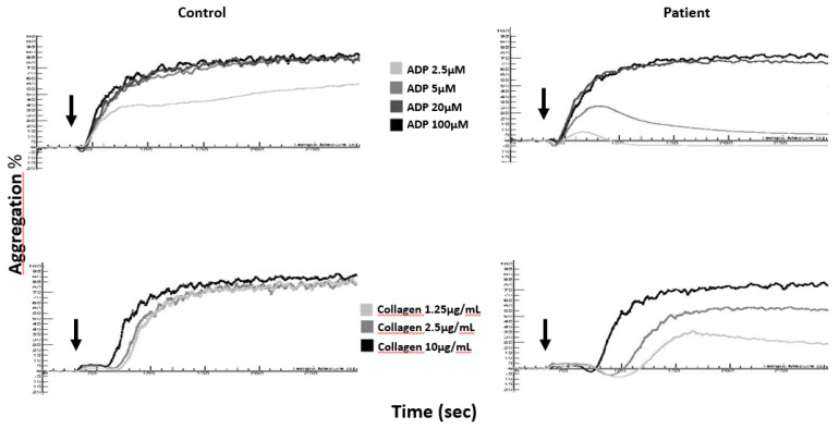
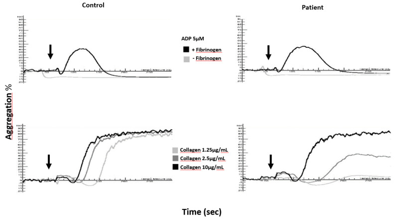
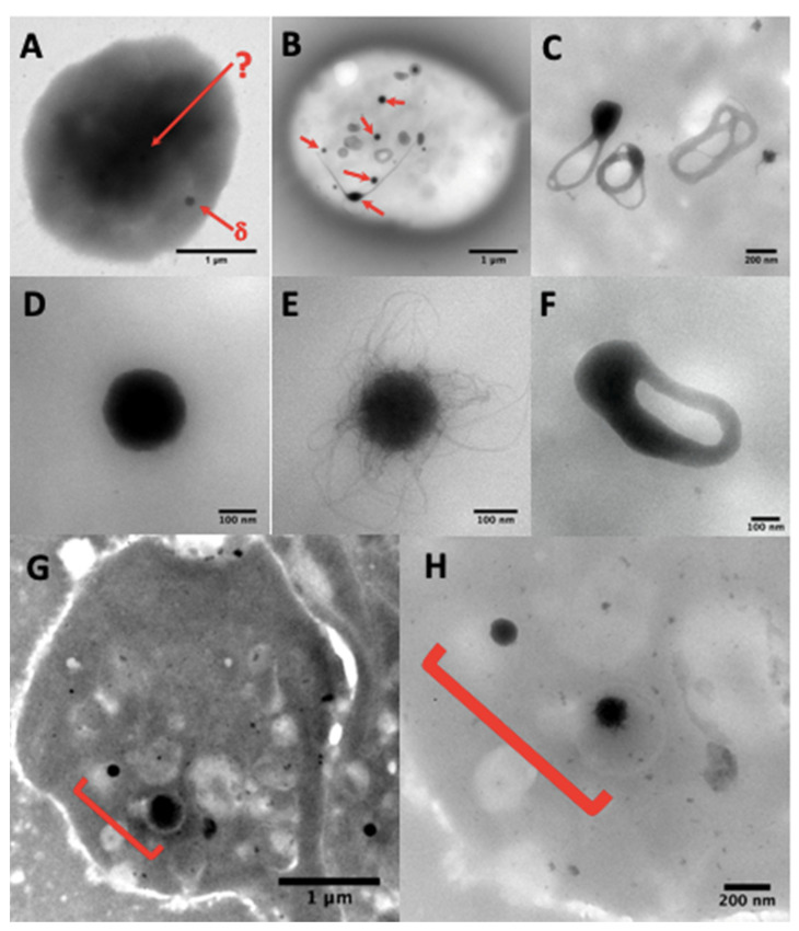
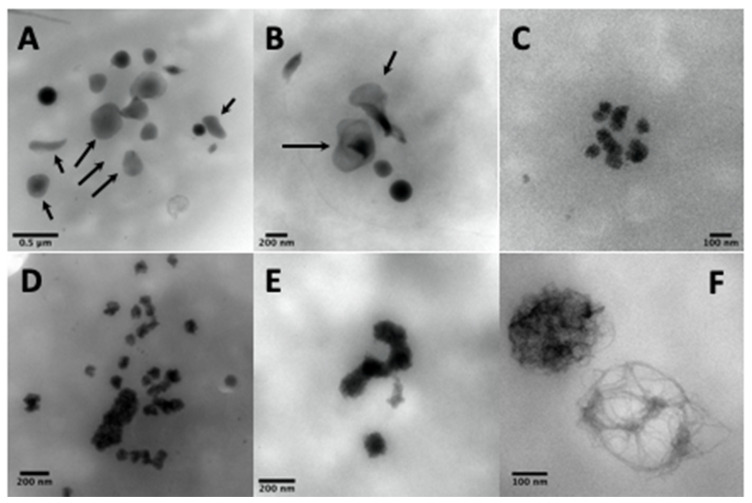
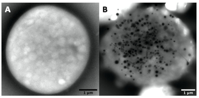
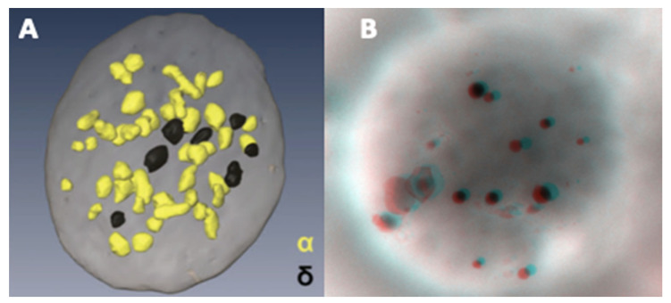
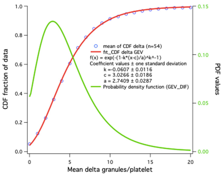
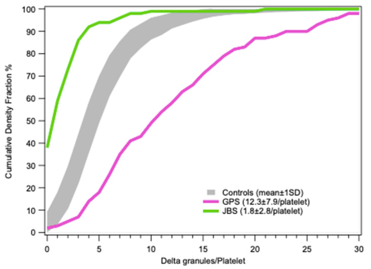
Platelet dense-granules are small organelles specific to the platelet lineage that contain small molecules (calcium, adenyl nucleotides, serotonin) and are essential for the activation of blood platelets prior to their aggregation in the event of a vascular injury. Delta-storage pool diseases (δ-SPDs) are platelet pathologies leading to hemorrhagic syndromes of variable severity and related to a qualitative (content) or quantitative (numerical) deficiency in dense-granules. These pathologies appear in a syndromic or non-syndromic form. The syndromic forms (Chediak-Higashi disease, Hermansky-Pudlak syndromes), whose causative genes are known, associate immune deficiencies and/or oculocutaneous albinism with a platelet function disorder (PFD). The non-syndromic forms correspond to an isolated PFD, but the genes responsible for the pathology are not yet known. The diagnosis of these pathologies is complex and poorly standardized. It is based on orientation tests performed by light transmission aggregometry or flow cytometry, which are supplemented by complementary tests based on the quantification of platelet dense-granules by electron microscopy using the whole platelet mount technique and the direct determination of granule contents (ADP/ATP and serotonin). The objective of this review is to present the state of our knowledge concerning platelet dense-granules and the tools available for the diagnosis of different forms of δ-SPD.

Keywords: blood platelets; electron microscopy; inherited platelet disorders; storage pool disorder.


Conflict of interest statement

The authors declare no conflict of interest.

Figures

Figure 1
Figure 1
Aggregation curves in citrated platelet-rich plasma (cPRP) of a δ-SPD patient and a healthy control (Control) in response to ADP (2.5, 5, 20, and 100 µM) and collagen (1.25, 2.5, and 10 µg/mL); arrows denote the times of addition of the agonists to cPRP.
Figure 2
Figure 2
Aggregation curves in a washed platelet suspension of a delta-storage pool disease (δ-SPD) patient and a healthy control (Control) in response to ADP (5 µM) and collagen (1.25, 2.5 and 10 µg/mL); arrows denote the times of addition of the agonists to washed platelets.
Figure 3
Figure 3
(A) A whole platelet mount from a platelet suspension incubated in 1.25% glutaraldehyde phosphate buffer for one hour. The use of aldehyde fixatives, even very briefly, is not recommended and paraformaldehyde treatment gives similar results; the platelet background becomes so dark that only some peripheral granules (δ-arrow) are sometimes visible, but no granules in the platelet center (δ-arrow). (B) dense-granules (red arrows). A simple brief wash in pure water of native platelets on a formvar film is sufficient to obtain electron-translucent cells where the dense-granules are perfectly identifiable. These granules are classically smooth (D) or with filaments (E), but are sometimes observable in restructuring phases as annular forms (C) that split into long extensions (B, bottom left center), possibly also in a division phase (F), where two nodules are visible, which will probably evolve into two well separated dense-granules. The camera in automatic contrast adjustment mode may make the identification of dense-granules doubtful. The quickest solution is to change the magnification rather than the long contrast-brightness-gamma settings; an element looking like a veil at Mag 20 k (G) will be better identifiable at Mag 80 k (H).
Figure 4
Figure 4
The electron-dense elements not counted as dense-granules are calcium- and phosphorus-poor veils [67] (A,B, arrows), clusters of spongy elements (C), also present in the form of chains or larger grains (D,E), which are found in abundance and bigger in STIM1–York–Stormorken pathologies and are rich in calcium and phosphorus [70], while others are poorer in calcium (F), which could represent immature dense-granules (possibly polyphosphate filaments without calcium accumulation, found in non-identified cases of delta deficiency, but not in Hermansky–Pudlak syndrome).
Figure 5
Figure 5
Examples of a platelet totally devoid of electron-dense material (A) like those found in Hermansky–Pudlak syndrome (HPS) patients and a macro-platelet of a gray platelet syndrome (GPS) patient (B) with 155 dense-granules (mean 14 ± 26/platelet, 17% of platelets with >20 dense-granules).
Figure 6
Figure 6
Using conventional electron microscopy with 70 nm thick sections, the probability of observing a dense granule is low. An alternative is the reconstruction of serial sections. The rapid, more, or less automated method of focused ion beam-scanning electron microscopy (FIB-SEM) makes it possible to observe the spatial distribution of α- (yellow) and dense-granules (black) in a platelet. (A) data from Eckly et al. published in [76] (part of Figure 6B, image obtained from the Haematologica Journal website http://www.haematologica.org). A stereo image of an entire platelet on formvar can be obtained from two tilt images taken at ±7°. (B) Anaglyph reconstructed with ImageJ (V1.8 NIH, USA) Two Shot Anaglyph software (V2.9.5, Sandy Knoll Software, USA); the stereo effect is visible with red-cyan glasses.
Figure 7
Figure 7
Distribution model of the number of dense-granules/platelet: cumulative distribution frequency (CDF) averages in 54 controls (blue circles). The simplest fit is a generalized extreme value (GEV) distribution function (red); the corresponding probability density obtained by CDF derivative (Igor Wavemetrics) is shown in green.
Figure 8
Figure 8
Cumulative frequencies of dense-granule distributions in platelets (mean ± 1SD) in controls (gray, n = 54) and two cases of macrothombocytopenia with similar platelet size distributions, a patient with GPS whose platelet dense granule content may be considered to be normal and a patient with Jacobsen syndrome (JBS) having a severe deficit.

References

    1. Versteeg H.H., Heemskerk J.W., Levi M., Reitsma P.H. New fundamentals in hemostasis. Physiol. Rev. 2013;93:327–358. doi: 10.1152/physrev.00016.2011. - DOI - PubMed
    1. Sharda A., Flaumenhaft R. The life cycle of platelet granules. F1000Res. 2018;7:236. doi: 10.12688/f1000research.13283.1. - DOI - PMC - PubMed
    1. Gachet C. Regulation of platelet functions by P2 receptors. Annu. Rev. Pharmacol. Toxicol. 2006;46:277–300. doi: 10.1146/annurev.pharmtox.46.120604.141207. - DOI - PubMed
    1. Masliah-Planchon J., Darnige L., Bellucci S. Molecular determinants of platelet delta storage pool deficiencies: An update. Br. J. Haematol. 2013;160:5–11. doi: 10.1111/bjh.12064. - DOI - PubMed
    1. Flaumenhaft R. Platelet Secretion—Chapter 18. In: Michelson A.D., editor. Platelets. 3rd ed. Academic Press; Cambridge, MA, USA: 2013. pp. 343–366. - DOI

LinkOut - more resources