Review
. 2020 Aug 4;9(8):705.
doi: 10.3390/antiox9080705.

Extracellular Vesicles and MicroRNA: Putative Role in Diagnosis and Treatment of Diabetic Retinopathy

Affiliations
Review

Extracellular Vesicles and MicroRNA: Putative Role in Diagnosis and Treatment of Diabetic Retinopathy

Beatriz Martins et al. Antioxidants (Basel). .

Abstract

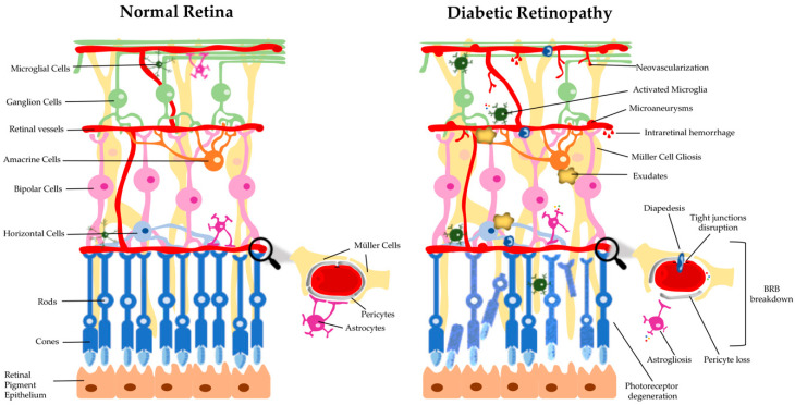
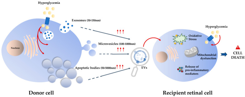
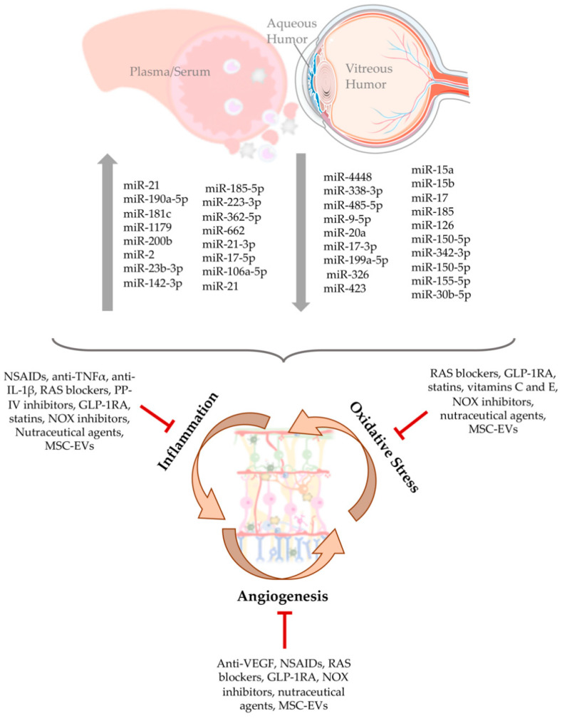
Diabetic retinopathy (DR) is a complex, progressive, and heterogenous retinal degenerative disease associated with diabetes duration. It is characterized by glial, neural, and microvascular dysfunction, being the blood-retinal barrier (BRB) breakdown a hallmark of the early stages. In advanced stages, there is formation of new blood vessels, which are fragile and prone to leaking. This disease, if left untreated, may result in severe vision loss and eventually legal blindness. Although there are some available treatment options for DR, most of them are targeted to the advanced stages of the disease, have some adverse effects, and many patients do not adequately respond to the treatment, which demands further research. Oxidative stress and low-grade inflammation are closely associated processes that play a critical role in the development of DR. Retinal cells communicate with each other or with another one, using cell junctions, adhesion contacts, and secreted soluble factors that can act in neighboring or long-distance cells. Another mechanism of cell communication is via secreted extracellular vesicles (EVs), through exchange of material. Here, we review the current knowledge on deregulation of cell-to-cell communication through EVs, discussing the changes in miRNA expression profiling in body fluids and their role in the development of DR. Thereafter, current and promising therapeutic agents for preventing the progression of DR will be discussed.

Keywords: angiogenesis; antioxidants; biomarkers; diabetic retinopathy (DR); extracellular vesicles; inflammation; miRNA; oxidative stress.


Conflict of interest statement

The authors declare no conflict of interest.

Figures

Figure 1
Figure 1
Schematic drawing of a cross-section of a healthy retina and a retina with diabetic retinopathy. The retina is a multilayered tissue composed by neuronal (photoreceptors, and horizontal, bipolar, amacrine and ganglion cells) and glial (astrocytes, Müller cells, and microglia) cells, that closely embed with three capillary plexuses. The blood-retinal barrier (BRB), that regulates the exchange of fluids between blood and retinal tissue, plays an important role in the maintenance of the retinal homeostasis. However, in diabetic retinopathy (DR) several retinal abnormalities appear, including microvascular changes (microaneurysms, intra-retinal hemorrhages, and neovascularization), the appearance of exudates, ganglion cell and photoreceptor degeneration, and glial dysfunction (astrogliosis, Müller cell gliosis, and activation of microglia). The reactivity of glial cells promotes the secretion of inflammatory cytokines, leukocyte adhesion, and diapedesis. These alterations lead to the breakdown of the BRB, characterized by pericyte loss and tight junctions’ disruption.
Figure 2
Figure 2
Extracellular vesicles (EVs) and their role in the pathogenesis of diabetic retinopathy (DR). EVs can be classified into three different types depending on their size and biogenesis—exosomes, microvesicles, and apoptotic bodies. Hyperglycemia leads to the increase of EVs released from donor cells. These EVs, derived from other retinal cells or from circulation, can interact with recipient/target retinal cells contributing, along with chronic hyperglycemia, to oxidative stress and release of pro-inflammatory mediators. These mechanisms may contribute to mitochondrial dysfunction leading to retinal cell death.
Figure 3
Figure 3
Alterations of miRNAs profile in DR and possible therapeutic interventions. The progression of DR is associated with the deregulation of miRNA expression, which leads to the increase (↑) or decrease (↓) of some miRNA involved in oxidative stress, inflammatory, and angiogenic processes. However, some therapeutic and nutraceutical strategies targeting inflammatory pathways, oxidative stress, and neovascularization, can be useful in preventing or arresting DR progression.

References

    1. Federation I.D. IDF Diabetes Atlas. 9th ed. International Diabetes Federation; Brussels, Belgium: 2019.
    1. Wang P., Chen F., Wang W., Zhang X.D. Hydrogen Sulfide Attenuates High Glucose-Induced Human Retinal Pigment Epithelial Cell Inflammation by Inhibiting ROS Formation and NLRP3 Inflammasome Activation. Mediat. Inflamm. 2019;2019:1–13. doi: 10.1155/2019/8908960. - DOI - PMC - PubMed
    1. Youngblood H., Robinson R., Sharma A., Sharma S. Proteomic Biomarkers of Retinal Inflammation in Diabetic Retinopathy. Int. J. Mol. Sci. 2019;20:4755. doi: 10.3390/ijms20194755. - DOI - PMC - PubMed
    1. Biswas S., Sarabusky M., Chakrabarti S. Diabetic Retinopathy, lncRNAs, and Inflammation: A Dynamic, Interconnected Network. J. Clin. Med. 2019;8:1033. doi: 10.3390/jcm8071033. - DOI - PMC - PubMed
    1. Petrie J.R., Guzik T.J., Touyz R.M. Diabetes, Hypertension, and Cardiovascular Disease: Clinical Insights and Vascular Mechanisms. Can. J. Cardiol. 2018;34:575–584. doi: 10.1016/j.cjca.2017.12.005. - DOI - PMC - PubMed

LinkOut - more resources