

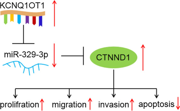
Long non-coding RNA KCNQ1OT1 up-regulates CTNND1 by sponging miR-329-3p to induce the proliferation, migration, invasion, and inhibit apoptosis of colorectal cancer cells
- PMID: 32760218
- PMCID: PMC7379774
- DOI: 10.1186/s12935-020-01425-2
Long non-coding RNA KCNQ1OT1 up-regulates CTNND1 by sponging miR-329-3p to induce the proliferation, migration, invasion, and inhibit apoptosis of colorectal cancer cells
Abstract
Background: Long non-coding RNAs (lncRNAs) have been certified to be involved in the occurrence and growth of diverse cancers, including CRC. The purpose of the research was to explore the effects of lncRNA KCNQ1 overlapping transcript 1 (KCNQ1OT1) on proliferation, migration, invasion, and apoptosis in CRC cells and its mechanism.
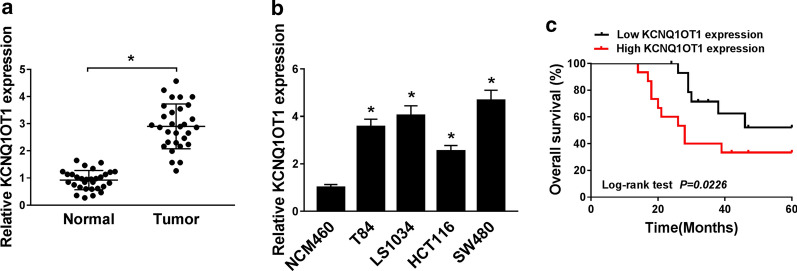
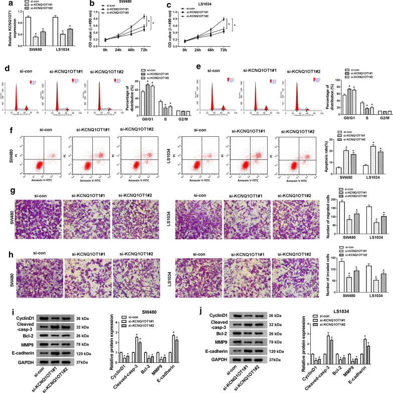
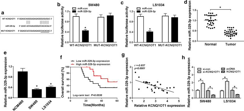
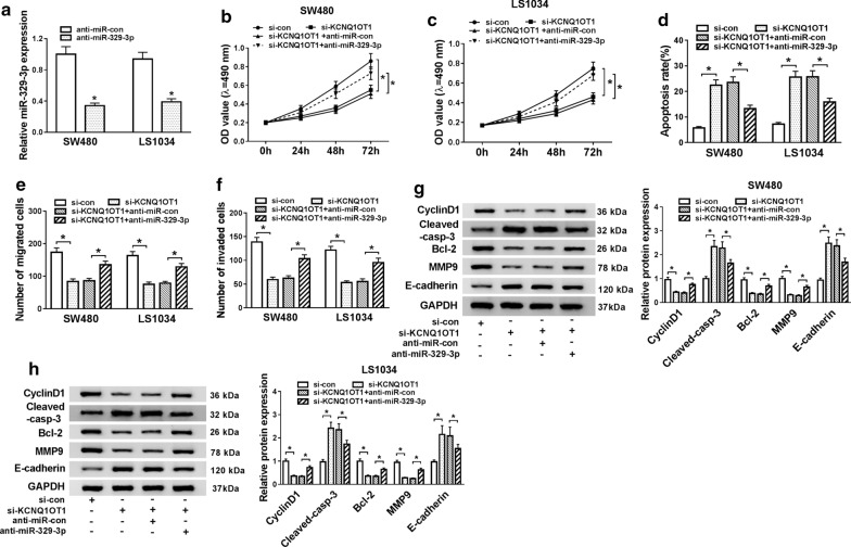
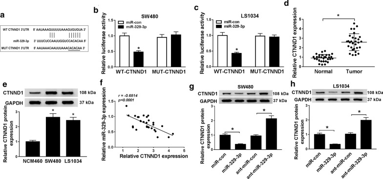
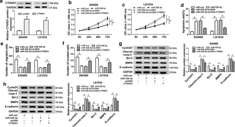
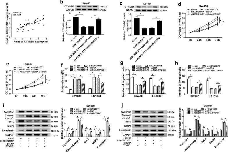
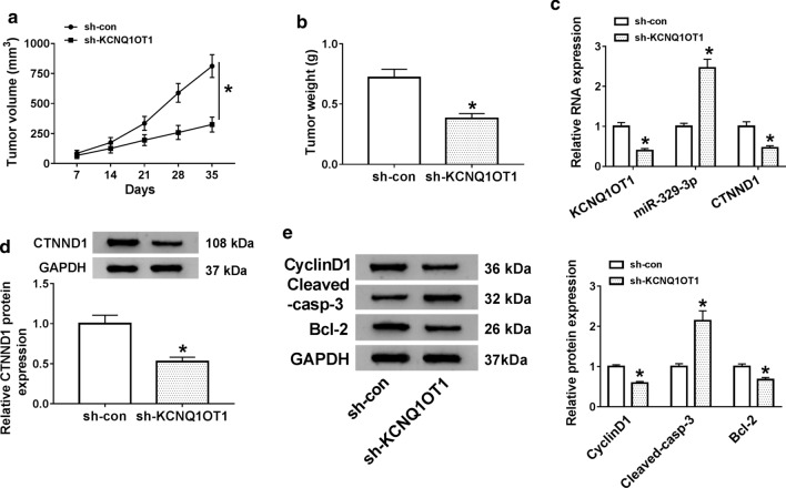
Methods: The levels of KCNQ1OT1 and miR-329-3p were examined by quantitative real-time polymerase chain reaction (qRT-PCR) in CRC tissues and cells. The mRNA and protein levels of catenin delta-1 (CTNND1) were measured by qRT-PCR and western blot analysis, respectively. The targets of KCNQ1OT1 and miR-329-3p were predicted by online software and confirmed by luciferase reporter assay. The cell proliferation, migration, invasion, and apoptosis were examined using 3-(4, 5-dimethylthiazol-2-yl)-2, 5-diphenyltetrazolium bromide (MTT), transwell, and apoptosis assay. The expression levels of CyclinD1, Bcl-2, MMP9, Cleaved-casp-3, and E-cadherin in SW480 and LS1034 cells were gauged by western blot analysis. Xenograft tumor model was structured to prove the biological role of KCNQ1OT1 of CRC in vivo.
Results: The levels of KCNQ1OT1 and CTNND1 were significantly increased in CRC tissues and cells. Knockdown of KCNQ1OT1 suppressed proliferation, migration, invasion, and induced apoptosis in CRC cells. Conversely, CTNND1 overexpression reversed the impact of KCNQ1OT1 knockdown on CRC cells. Moreover, CTNND1 was verified as a direct target of miR-329-3p, and miR-329-3p could specially bind to KCNQ1OT1. Also, the down-regulation of KCNQ1OT1 triggered the CRC progress by up-regulating CTNND1 expression in CRC cells. Besides, KCNQ1OT1 knockdown inhibited CRC tumor growth through the miR-329-3p/CTNND1 axis in vivo.
Conclusion: Our results indicated that KCNQ1OT1 could positively regulate CTNND1 expression by sponging miR-329-3p, thereby boosting the progression of CRC. Our findings provided the underlying therapy targets for CRC.
Keywords: CTNND1; Colorectal cancer; lncRNA KCNQ1OT1; miR-329-3p.
© The Author(s) 2020.
Conflict of interest statement
Competing interestsThe authors declare that there are no competing interests.
Figures
References
LinkOut - more resources
Full Text Sources
Research Materials
Miscellaneous
