

11 C-PK11195 PET-based molecular study of microglia activation in SOD1 amyotrophic lateral sclerosis
- PMID: 32762033
- PMCID: PMC7480909
- DOI: 10.1002/acn3.51112
11 C-PK11195 PET-based molecular study of microglia activation in SOD1 amyotrophic lateral sclerosis
Abstract
Objective: Neuroinflammation is considered a key driver for neurodegeneration in several neurological diseases, including amyotrophic lateral sclerosis (ALS). SOD1 mutations cause about 20% of familial ALS, and related pathology might generate microglial activation triggering neurodegeneration. 11 C-PK11195 is the prototypical and most validated PET radiotracer, targeting the 18-kDa translocator protein which is overexpressed in activated microglia. In this study, we investigated microglia activation in asymptomatic (ASYM) and symptomatic (SYM) SOD1 mutated carriers, by using 11 C-PK11195 and PET imaging.
Methods: We included 20 subjects: 4 ASYM-carriers, neurologically normal, 6 SYM-carriers with probable ALS, and 10 healthy controls. A receptor parametric mapping procedure estimated 11 C-PK11195 binding potentials and voxel-wise statistical comparisons were performed at group and single-subject levels.
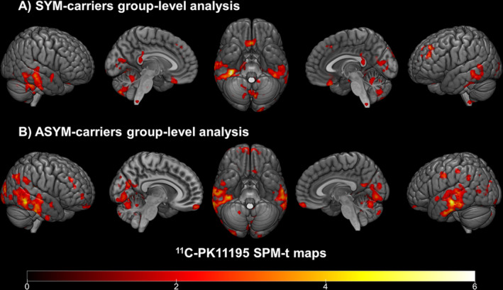
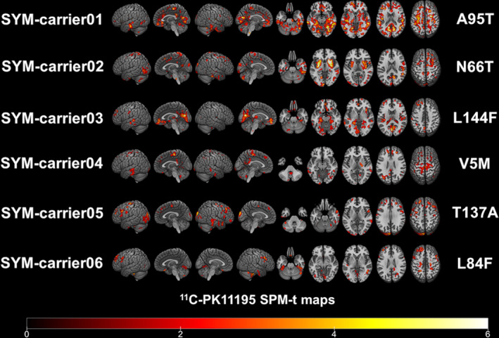
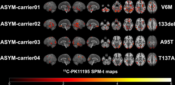
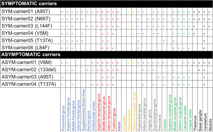
Results: Both the SYM- and ASYM-carriers showed significant microglia activation in cortical and subcortical structures, with variable patterns at individual level. Clusters of activation were present in occipital and temporal regions, cerebellum, thalamus, and medulla oblongata. Notably, SYM-carriers showed microglia activation also in supplementary and primary motor cortices and in the somatosensory regions.
Interpretation: In vivo neuroinflammation occurred in all SOD1 mutated cases since the presymptomatic stages, as shown by a significant cortical and subcortical microglia activation. The involvement of sensorimotor cortex became evident at the symptomatic disease stage. Although our data indicate the role of in vivo PET imaging for assessing resident microglia in the investigation of SOD1-ALS pathophysiology, further studies are needed to clarify the temporal and spatial dynamics of microglia activation and its relationship with neurodegeneration.
© 2020 The Authors. Annals of Clinical and Translational Neurology published by Wiley Periodicals LLC on behalf of American Neurological Association.
Conflict of interest statement
The authors declare that they have no conflict of interest relevant for this article.
Figures
References
-
- Robberecht W., Philips T.. The changing scene of amyotrophic lateral sclerosis. Nat Rev Neurosci 2013;14:248–264. - PubMed
-
- Kiernan M.C., Vucic S., Cheah B.C., et al. Amyotrophic lateral sclerosis. Lancet 2011;377:942–955. - PubMed
-
- De Marchi F., Sarnelli M.F., Solara V., et al. Depression and risk of cognitive dysfunctions in amyotrophic lateral sclerosis. Acta Neurol Scand 2019;139:438–445. - PubMed
-
- Chiò A., Mazzini L., Mora G.. Disease‐modifying therapies in amyotrophic lateral sclerosis. Neuropharmacology 2020;107986. - PubMed
Publication types
MeSH terms
Substances
LinkOut - more resources
Full Text Sources
Medical
Miscellaneous
