Review
. 2020 Nov;18(11):2812-2822.
doi: 10.1111/jth.15050. Epub 2020 Sep 18.

Is the COVID-19 thrombotic catastrophe complement-connected?

Affiliations
Review

Is the COVID-19 thrombotic catastrophe complement-connected?

Edward M Conway et al. J Thromb Haemost. 2020 Nov.

Abstract

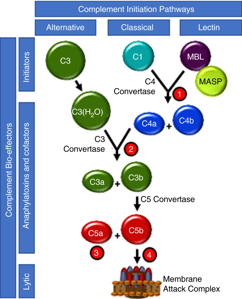
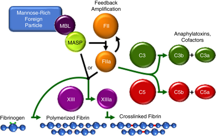
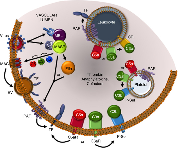
In December 2019, the world was introduced to a new betacoronavirus, referred to as severe acute respiratory syndrome coronavirus 2 (SARS-CoV-2) for its propensity to cause rapidly progressive lung damage, resulting in high death rates. As fast as the virus spread, it became evident that the novel coronavirus causes a multisystem disease (COVID-19) that may involve multiple organs and has a high risk of thrombosis associated with striking elevations in pro-inflammatory cytokines, D-dimer, and fibrinogen, but without disseminated intravascular coagulation. Postmortem studies have confirmed the high incidence of venous thromboembolism, but also notably revealed diffuse microvascular thrombi with endothelial swelling, consistent with a thrombotic microangiopathy, and inter-alveolar endothelial deposits of complement activation fragments. The clinicopathologic presentation of COVID-19 thus parallels that of other thrombotic diseases, such as atypical hemolytic uremic syndrome (aHUS), that are caused by dysregulation of the complement system. This raises the specter that many of the thrombotic complications arising from SARS-CoV-2 infections may be triggered and/or exacerbated by excess complement activation. This is of major potential clinical relevance, as currently available anti-complement therapies that are highly effective in protecting against thrombosis in aHUS, could be efficacious in COVID-19. In this review, we provide mounting evidence for complement participating in the pathophysiology underlying the thrombotic diathesis associated with pathogenic coronaviruses, including SARS-CoV-2. Based on current knowledge of complement, coagulation and the virus, we suggest lines of study to identify novel therapeutic targets and the rationale for clinical trials with currently available anti-complement agents for COVID-19.

Keywords: complement; covid-19; microvascular; thrombotic microangiopathy; tissue factor.


Figures

FIGURE 1
FIGURE 1
Complement pathway overview and therapeutic intervention. The three pathways of complement initiation are depicted. The alternative pathway (AP) is initiated by spontaneous hydrolysis of the intramolecular thioester within C3, thus providing a continuous source of the C3b‐like cofactor C3(H2O) that accelerates the proteolytic generation of the cofactor C3b for pathway propagation and anaphylatoxin C3a generation. The classical pathway (CP) is initiated by association of the C1q/r/s complex with antibody bound to a multivalent foreign particle, resulting in generation of the cofactor C4b, which accelerates enzymatic generation of C3a and C3b and mild anaphylatoxin C4a. Like the CP, initiation of the lectin pathway (LP) by recognition of foreign lectins by the MBL/MASP complex results in C3b/C3a production via C3 convertase. Once enough C3b accumulates, its cofactor activity produces the cofactor C5b, which triggers MAC assembly and foreign membrane permeabilization. The most potent complement anaphylatoxin, C5a, is a by‐product. Several complement loci have been targeted for therapeutic intervention: ①, CP/LP pathway initiation is inhibited by the serpin, CI‐INH, at the level of MASP and C1r; ②, C3a and C3b product formation is inhibited by the compstatin derivatives AMY‐101 and APL‐2; ③, the anaphylatoxin C5a interaction with its cellular receptor is prevented by the humanized monoclonal antibody, BDB001; and ④ C5b and C5a generation is inhibited by the humanized monoclonal antibodies, eculizumab and ravulizumab, thus preventing MAC assembly
FIGURE 2
FIGURE 2
The humoral MASP‐thrombin coagulation amplification cycle and reciprocal thrombin‐complement crossover. When associated with a mannose‐rich foreign particle (such as a virus‐infected cell surface), MBL triggers MASP‐mediated prothrombin (FII) activation to thrombin (FIIa), which feedback amplifies its own generation via the coagulation cascade (black lines). MASP and thrombin share substrate specificity resulting in cell modulation and crosslink‐stabilized clot formation. Thrombin crosses over into complement by cleaving C3 and C5, propagating inflammation and anaphylaxis
FIGURE 3
FIGURE 3
Complement‐ and coagulation‐mediated cell activation in COVID‐19. SARS‐CoV‐2‐encoded spike (SP) and nucleocapsid (NP) proteins can directly induce LP stimulation via MBL and MASP, respectively, triggering the complement‐coagulation crossover pathways and deposition of membrane attack complex (MAC) on infected cell surfaces. Perturbation of the cell membrane by MAC is known to induce extracellular vesicles (EV), which contain TF, thus further amplifying thrombin generation. Thrombin and MASP directly cleave protease activated receptors (PAR) leading to activation of endothelial cells, leukocytes, and platelets and additional production of thrombin. Ligand engagement by C5a (C5aR) and C3a (C3aR) receptors induces numerous cell changes including P‐selectin (P‐Sel) stimulation, which can localize complement cofactors C3b and C3(H2O) on the endothelium and platelets. C5aR and C3aR on leukocytes also trigger activation of complement receptors (CR), which bind C3b and its degradation products that modulate cell function. The stimulation of anaphylatoxin receptors and PARs can cause release of pro‐inflammatory intracellular granule contents. Thus, virus‐induced provocation of complement cofactors and anaphylatoxins and thrombin, cause cellular amplification of thromboinflammation

References

    1. Zhu N., Zhang D., Wang W., et al. A novel coronavirus from patients with pneumonia in China, 2019. N Engl J Med. 2020;382(8):727–733. - PMC - PubMed
    1. Zhou F., Yu T., Du R., et al. Clinical course and risk factors for mortality of adult inpatients with COVID‐19 in Wuhan, China: a retrospective cohort study. Lancet. 2020;395(10229):1054–1062. - PMC - PubMed
    1. Giannis D., Ziogas I.A., Gianni P. Coagulation disorders in coronavirus infected patients: COVID‐19, SARS‐CoV‐1, MERS‐CoV and lessons from the past. J Clin Virol. 2020;127:104362. - PMC - PubMed
    1. Lodigiani C., Iapichino G., Carenzo L., et al. Venous and arterial thromboembolic complications in COVID‐19 patients admitted to an academic hospital in Milan, Italy. Thromb Res. 2020;191:9–14. - PMC - PubMed
    1. Klok F.A., Kruip M., van der Meer N.J.M., et al. Confirmation of the high cumulative incidence of thrombotic complications in critically ill ICU patients with COVID‐19: an updated analysis. Thromb Res. 2020;19:148–150. - PMC - PubMed

Publication types

Substances