

Case Reports
doi: 10.1111/bpa.12889.
Epub 2020 Aug 28.
Neuropathologic features of four autopsied COVID-19 patients
Affiliations
- PMID: 32762083
- PMCID: PMC7436498
- DOI: 10.1111/bpa.12889
Item in Clipboard
Case Reports
Neuropathologic features of four autopsied COVID-19 patients
Brain Pathol.
2020 Nov.
No abstract available
Conflict of interest statement
The authors declare no conflict of interest.
Figures
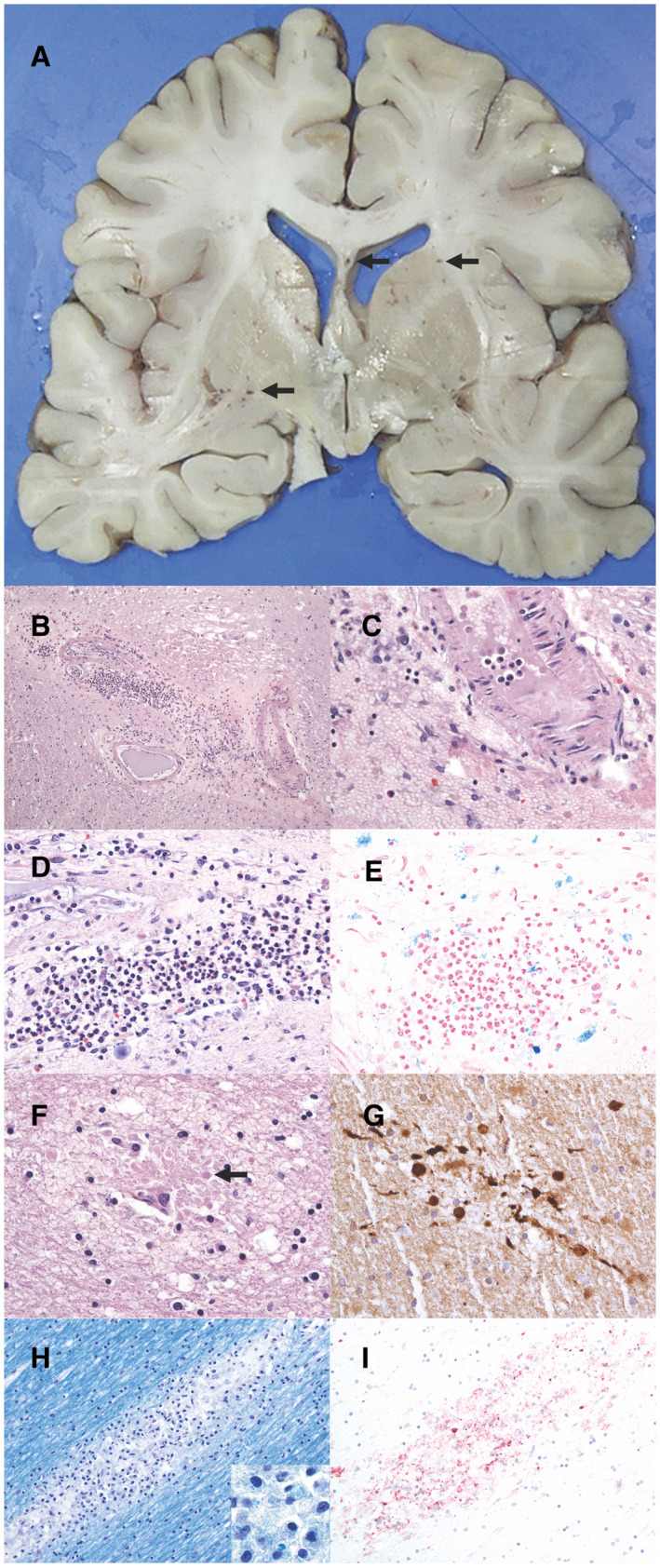
A. Coronal section of cerebral hemispheres exhibiting abundant enlarged perivascular spaces and microhemorrhages in the white matter and deep gray matter (arrows). In inferior putamen (both sides) lacunae are seen. B. Microscopic section from the internal capsule region shows abundant perivascular inflammation and perivascular hemorrhage, but very few inflammatory cells in the surrounding brain areas (H&E, 40× magnification). C–E. Higher resolution images from the region exhibited in B (H&E, 400× magnification). In (C), a small amount of fibrinoid material is seen in the lumen of the vessel. D. Abundant granulocytes are detected in this region in addition to perivascular hemorrhage. E. Iron staining of the same location as in (D) shows faint positivity. F. A small focus of perivascular hemorrhage, with axonal edema and spheroids (arrow) (H&E, 400× magnification), highlighted by IHC for APP in (G) (400× magnification). H. Luxol fast blue staining from the white matter of the cingular gyrus shows loss of myelin and microglial reaction (200× magnification). Macrophages engulfing myelin are shown in insert (1000× magnification). H&E staining of this section exhibited a vessel in this location (see Supporting Information). I. Iba‐1 IHC staining of the same location highlights the microglial cells (200× magnification).
References
Publication types
MeSH terms
Grants and funding
LinkOut - more resources
Full Text Sources
Medical
