. 2021 Feb;31(2):315-326.
doi: 10.1089/thy.2020.0332. Epub 2020 Sep 25.

Thyroid Hormone Receptor α Mutations Cause Heart Defects in Zebrafish

Affiliations

Thyroid Hormone Receptor α Mutations Cause Heart Defects in Zebrafish

Cho Rong Han et al. Thyroid. 2021 Feb.

Abstract

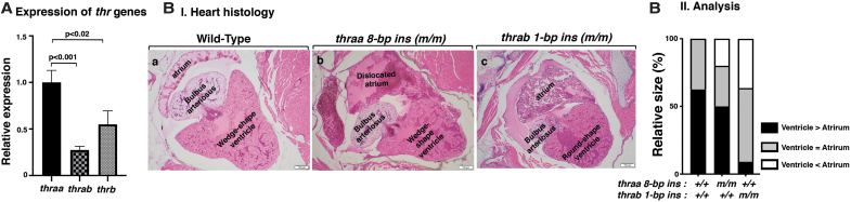
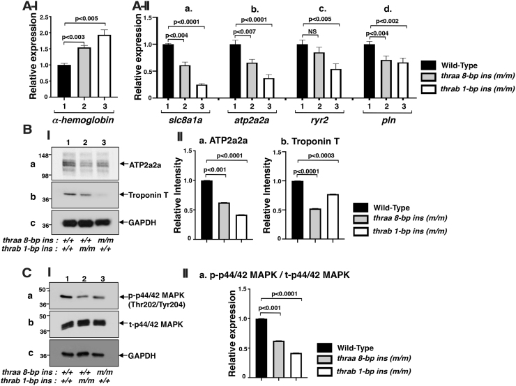
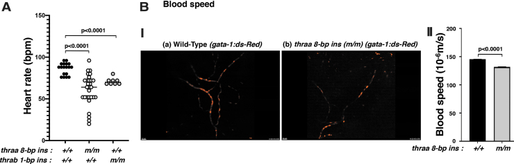
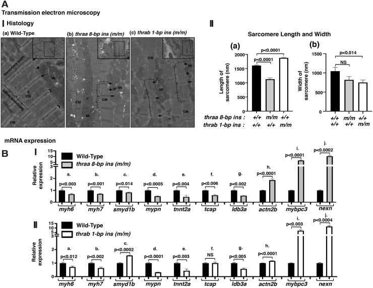
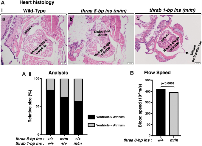
Background: Mutations of thyroid hormone receptor α1 (TRα1) cause resistance to thyroid hormone (RTHα). Patients exhibit growth retardation, delayed bone development, anemia, and bradycardia. By using mouse models of RTHα, much has been learned about the molecular actions of TRα1 mutants that underlie these abnormalities in adults. Using zebrafish models of RTHα that we have recently created, we aimed to understand how TRα1 mutants affect the heart function during this period. Methods: In contrast to human and mice, the thra gene is duplicated, thraa and thrab, in zebrafish. Using CRISPR/Cas9-mediated targeted mutagenesis, we created C-terminal mutations in each of two duplicated thra genes in zebrafish (thraa 8-bp insertion or thrab 1-bp insertion mutations). We recently showed that these mutant fish faithfully recapitulated growth retardation as found in patients and thra mutant mice. In the present study, we used histological analysis, gene expression profiles, confocal fluorescence, and transmission electron microscopy (TEM) to comprehensively analyze the phenotypic characteristics of mutant fish heart during development. Results: We found both a dilated atrium and an abnormally shaped ventricle in adult mutant fish. The retention of red blood cells in the two abnormal heart chambers, and the decreased circulating blood speed and reduced expression of contractile genes indicated weakened contractility in the heart of mutant fish. These abnormalities were detected in mutant fish as early as 35 days postfertilization (juveniles). Furthermore, the expression of genes associated with the sarcomere assembly was suppressed in the heart of mutant fish, resulting in abnormalities of sarcomere organization as revealed by TEM, suggesting that the abnormal sarcomere organization could underlie the bradycardia exhibited in mutant fish. Conclusions: Using a zebrafish model of RTHα, the present study demonstrated for the first time that TRα1 mutants could act to cause abnormal heart structure, weaken contractility, and disrupt sarcomere organization that affect heart functions. These findings provide new insights into the bradycardia found in RTHα patients.

Keywords: blood speed; bradycardia; contractility; heart defects; mutations; zebrafish, TRα.


Conflict of interest statement

No competing financial interests exist.

Figures

FIG. 1.
FIG. 1.
The mutation of thra genes results in cardiac atrial dilation in adult homozygous thra (m/m) mutant zebrafish. (A) The mRNA expression of thyroid hormone receptor genes (thraa, thrab, and thrb) in the heart of WT fish (3.7 mpf) was determined by RT-PCR as described in the Materials and Methods section. The number of WT fish analyzed is 14 (N = 14). The data are shown as mean ± SE (n = 3, three biological samples, each triplicate), with p-values to indicate significant changes. Two-tailed unpaired t-test, p-adjusted <0.05, was used for statistical analysis. GraphPad Prism version 8 for Mac OS X was used to perform analyses of variances. (B[I]) Representative histology features of zebrafish heart in WT (4 mpf) (B[a]); homozygous thraa 8-bp ins (m/m) (4.1 mpf) (B[b]); and homozygous thrab 1-bp ins (m/m) (5.1 mpf) (B[c]), sectioned and stained with H&E. The histology features of heart in thraa 8-bp ins (m/m) mutant fish show laterally displaced and dilated atrium; thrab 1-bp ins (m/m) mutant fish show a round-shaped ventricle and dilated atrium. (B[II]) Semiquantitative analysis of relative size of ventricle to atrium. Relative ventricle and atrium sizes at the aortic valve and atrioventricular valves are compared between WT (N = 6), thraa 8-bp ins (m/m) (N = 6), and thrab 1-bp ins (m/m) (N = 6) mutant fish. mpf, months postfertilization; RT-PCR, real-time polymerase chain reaction; SE, standard error; H&E, hematoxylin and eosin; WT, wild type.
FIG. 2.
FIG. 2.
Increased gene expression of α-hemoglobin (A[I]) altered calcium handling regulator gene mRNA expression (A[II]), protein abundance (B), and attenuated ERK signaling (C) in adult homozygous thra (m/m) mutant zebrafish. (A[I]) The mRNA expression of α-hemoglobin gene in the heart of WT, homozygous thraa 8-bp ins (m/m), and homozygous thrab 1-bp ins (m/m) fish (6.2 mpf) (N = 15). (A[II]) The mRNA expression of Ca2+ handling regulator genes, (a) slc8a1a, (b) atp2a2a, (c) ryr2, and (d) pln, in the heart of WT, homozygous thraa 8-bp ins (m/m), and homozygous thrab 1-bp ins (m/m) fish (11–12 mpf) (N = 10–12) was determined by RT-PCR as described in the Materials and Methods section. (B[I]) Western blot analysis was carried out for (a) ATP2a2a, (b) troponin T, and (c) GAPDH using heart as described in the Materials and Methods section. (B[II]) Quantitative analysis of relative protein abundance of the ratios of (a) ATP2a2a and (b) troponin T using GAPDH as a loading control. (C[I]) Western blot analysis of (a) p-ERK, (b) total ERK, and (c) GAPDH from the heart of fish with genotypes indicated. (C[II]) Quantitative analysis of relative protein abundance of the ratio of p-ERK to total ERK using GAPDH as a loading control. For Western blot analysis, the number of fish used was 9–11 (11 mpf). The data are shown as mean ± SE (n = 3) with p-values to indicate significant changes. Two-tailed unpaired t-test, p-adjusted <0.05, was used for statistical analysis. GraphPad Prism version 8 for Mac OS X was used to perform analyses of variances. NS, not significant.
FIG. 3.
FIG. 3.
thraa 8-bp ins (m/m) mutants show decreased heart rate (A) and decreased blood flow speed (B). (A) Heart rates in adult WT, thraa 8-bp ins (m/m), and thrab 1-bp ins (m/m) zebrafish were counted as described in the Materials and Methods section. For the measuring of heart rates, 8–29 fish (11–14 mpf) were used. (B[I]) Decreased flow speed of red blood cells in tg (thraa 8-bp ins(m/m):gata-1-dsRed) (b) compared with tg (WT: thraa+/+:gata-1-dsRed) (a) (2 mpf). The video can be viewed in Supplementary Figure S1. (B[II]) Comparison of the flow speed of red blood cells in WT and homozygous thraa 8-bp ins mutant fish. The images were captured from 3 to 4 individual WT or mutant fish; each fish was imagined 4–5 times. The data are shown as mean ± SE with p-values to indicate significant changes. Two-tailed unpaired t-test, p-adjusted <0.05, was used for statistical analysis. GraphPad Prism version 8 for Mac OS X was used to perform analyses of variances.
FIG. 4.
FIG. 4.
TEM shows myofilaments in ventricular sarcomere of adult zebrafish (A[I]). Representative micrographs of WT (a) and thraa 8-bp ins (m/m) (b) and thrab 1-bp ins (m/m) (c) mutants. Small lined rectangle indicates electron micrographs ( × 2000). Dotted rectangle areas were enlarged from each electron micrograph. Disrupted structure of ventricular sarcomere (length, width, and I-band) observed in thraa 8-bp ins (m/m) (b) and thrab 1-bp ins (m/m) (c) mutants compared with WT (a). Ventricular myocardium constituted of four to five overlapping layers of cardiac myocytes. Arrows point to Z-disc, crescent indicates I-band, arrow heads point to M-band, and brackets indicate representative sarcomere length (solid lines) and width (dotted lines), Mt; mitochondria, CM, cardiac myocytes. (A[II]). The length (a) and width (b) of individual sarcomeres were measured and graphed (n = 20) in randomly picked TEM images, the number of fish (N = 4 each genotype). The data are expressed as mean ± SE; the p-values are indicated. Two-tailed unpaired t-test, p-adjusted <0.05, was used for statistical analysis. (B) The mRNA expression of filaments and sarcomere structure genes in the heart of homozygous thraa 8-bp ins mutant fish (I) and homozygous thrab 1-bp ins (m/m) fish (11–12 mpf) (N = 10–12) (II) was determined by RT-PCR as described in the Materials and Methods section. The genes are as follows: (B[Ia] and B[IIa]) myh6, (B[Ib] and B[IIb]) myh7, (B[Ic] and B[IIc]) smyd1b, (B[Id] and B[IId]) mypn, (B[Ie] and B[IIe]) tnnt2, (B[If] and B[IIf]) tcap, (B[Ig] and B[IIg]) ldb3a, (B[Ih] and B[IIh]) actn2b, (B[Ii] and B[IIi]) mybpc3, and (B[Ij] and B[IIj]) nexn. The data are shown as mean ± SE (n = 3 biological samples, each with triplicates) with p-values to indicate significant changes. Two-tailed unpaired t-test, p-adjusted <0.05, was used for statistical analysis. GraphPad Prism version 8 for Mac OS X was used to perform analyses of variances. TEM, transmission electron microscopy.
FIG. 5.
FIG. 5.
Juveniles of homozygous thra mutant fish develop heart abnormalities. (A[I]) The histology features of zebrafish heart in WT (I[a]), homozygous thraa 8-bp ins (m/m) (I[b]), and homozygous thrab 1-bp ins (m/m) (I[c]) were sectioned and stained with H&E. The histology features of heart in thraa 8-bp ins (m/m) mutant fish show a dislocated and dilated atrium, and thrab 1-bp ins (m/m) mutant fish show a round-shaped ventricle and dilated atrium. (A[II]) Quantitative analysis of relative size of ventricle to atrium. Relative ventricle and atrium sizes at the aortic valve and atrioventricular valves are compared between WT (N = 3), thraa 8-bp ins (m/m) (N = 3), and thrab 1-bp ins (m/m) (N = 3) (1.1 mpf) mutant fish. GraphPad Prism version 8 for Mac OS X was used to perform analyses of variances. (B). Comparison of the flow speed of red blood cells in WT and homozygous thraa 8-bp ins mutant fish. The images were captured from 3 to 4 individual WT or mutant fish; each fish was imagined 4–5 times. Two-tailed unpaired t-test, p-adjusted <0.05, was used for statistical analysis. GraphPad Prism version 8 for Mac OS X was used to perform analyses of variances.

References

    1. Koenig RJ 1998. Thyroid hormone receptor coactivators and corepressors. Thyroid 8:703–713 - PubMed
    1. Kim MK, Lee JS, Chung JH. 1999. In vivo transcription factor recruitment during thyroid hormone receptor-mediated activation. Proc Natl Acad Sci U S A 96:10092–10097 - PMC - PubMed
    1. Vella KR, Ramadoss P, Costa ESRH, Astapova I, Ye FD, Holtz KA, Harris JC, Hollenberg AN. 2014. Thyroid hormone signaling in vivo requires a balance between coactivators and corepressors. Mol Cell Biol 34:1564–1575 - PMC - PubMed
    1. Ortiga-Carvalho TM, Sidhaye AR, Wondisford FE. 2014. Thyroid hormone receptors and resistance to thyroid hormone disorders. Nat Rev Endocrinol 10:582–591 - PMC - PubMed
    1. Mendoza A, Hollenberg AN. 2017. New insights into thyroid hormone action. Pharmacol Ther 173:135–145 - PMC - PubMed

Publication types

MeSH terms

Substances

LinkOut - more resources