Review
. 2020 Sep 1;319(3):H661-H681.
doi: 10.1152/ajpheart.00202.2020. Epub 2020 Aug 7.

Vascular mechanisms and molecular targets in hypertensive pregnancy and preeclampsia

Affiliations
Review

Vascular mechanisms and molecular targets in hypertensive pregnancy and preeclampsia

Hongmei Qu et al. Am J Physiol Heart Circ Physiol. .

Abstract

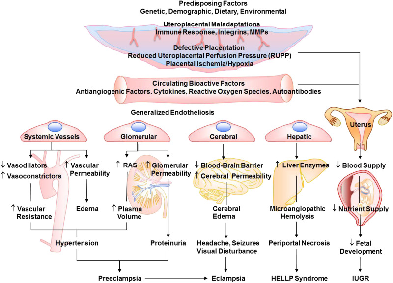
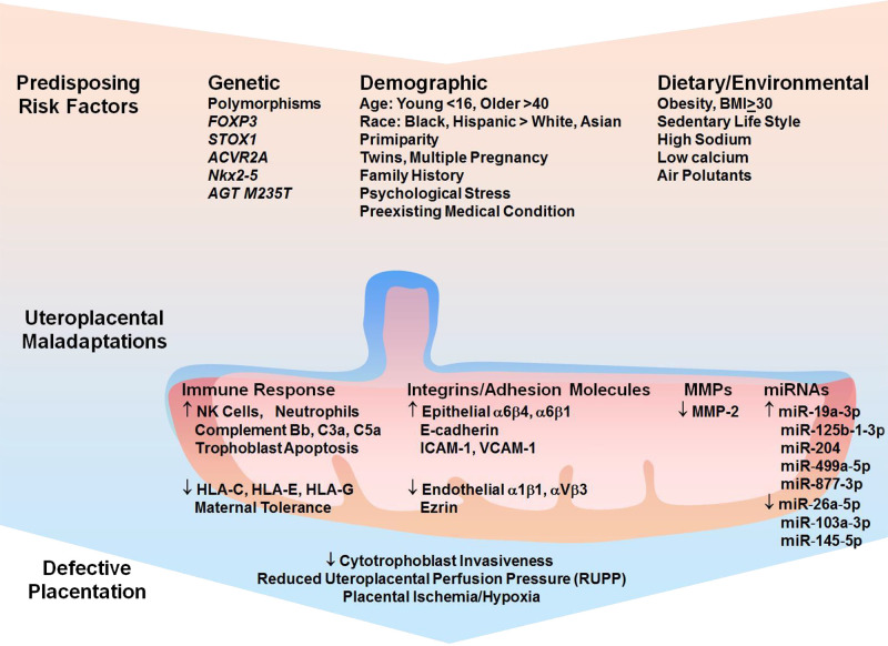
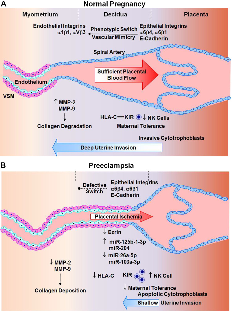
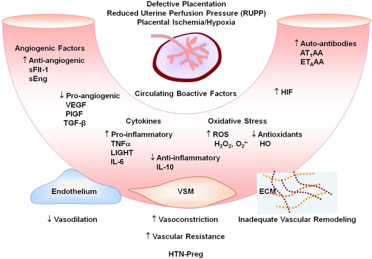
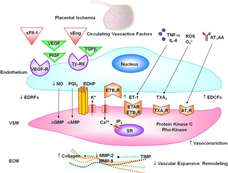
Preeclampsia is a major complication of pregnancy manifested as hypertension and often intrauterine growth restriction, but the underlying pathophysiological mechanisms are unclear. Predisposing genetic and environmental factors cause placental maladaptations leading to defective placentation, apoptosis of invasive cytotrophoblasts, inadequate expansive remodeling of the spiral arteries, reduced uteroplacental perfusion pressure, and placental ischemia. Placental ischemia promotes the release of bioactive factors into the maternal circulation, causing an imbalance between antiangiogenic soluble fms-like tyrosine kinase-1 and soluble endoglin and proangiogenic vascular endothelial growth factor, placental growth factor, and transforming growth factor-β. Placental ischemia also stimulates the release of proinflammatory cytokines, hypoxia-inducible factor, reactive oxygen species, and angiotensin type 1 receptor agonistic autoantibodies. These circulating factors target the vascular endothelium, causing generalized endotheliosis in systemic, renal, cerebral, and hepatic vessels, leading to decreases in endothelium-derived vasodilators such as nitric oxide, prostacyclin, and hyperpolarization factor and increases in vasoconstrictors such as endothelin-1 and thromboxane A2. The bioactive factors also target vascular smooth muscle and enhance the mechanisms of vascular contraction, including cytosolic Ca2+, protein kinase C, and Rho-kinase. The bioactive factors could also target matrix metalloproteinases and the extracellular matrix, causing inadequate vascular remodeling, increased arterial stiffening, and further increases in vascular resistance and hypertension. As therapeutic options are limited, understanding the underlying vascular mechanisms and molecular targets should help design new tools for the detection and management of hypertension in pregnancy and preeclampsia.

Keywords: cytokines; endothelium; hypertension; placental ischemia; vascular smooth muscle.


Conflict of interest statement

No conflicts of interest, financial or otherwise, are declared by the authors.

Figures

Fig. 1.
Fig. 1.
Risk factors, mechanisms, and manifestations of preeclampsia-related disorders. Predisposing risk factors cause placental maladaptations, placental ischemia, and the release of bioactive factors. Circulating bioactive factors cause generalized endotheliosis in systemic vessels leading to increased vascular resistance and hypertension, renal vessels leading to increased plasma volume and proteinuria, cerebral vessels leading to cerebral edema and neurological manifestations, and hepatic vessels leading to hemolysis, elevated liver enzymes, and low platelet count (HELLP) syndrome. Placental maladaptations and bioactive factors also reduce uteroplacental blood flow and lead to intrauterine growth restriction (IUGR). MMPs, matrix metalloproteinases; RAS, renin-angiotensin system.
Fig. 2.
Fig. 2.
Risk factors, uteroplacental maladaptations, and placental ischemia. Predisposing genetic polymorphisms, demographic characteristics, obesity, sedentary lifestyle, high-sodium low-calcium diet, and environmental pollutants could cause maladaptations in the uteroplacental immune response, integrins and adhesion molecules, matrix metalloproteinases (MMPs), and microRNAs. The uteroplacental maladaptations in turn lead to defective placentation and placental ischemia/hypoxia. BMI, body mass index; HLA-C, -E, and -G, histocompatibility complex molecules; NK, natural killer.
Fig. 3.
Fig. 3.
Defective placentation in preeclampsia. A: during normal pregnancy, phenotypic switch from epithelial to endothelial integrins, increased matrix metalloproteinase (MMP) expression/activity, and interaction of histocompatibility complex molecule HLA-C with its receptor rectifying K+ channel (KIR) and subsequent decrease in natural killer (NK) cell activity cause enhanced trophoblast invasion of spiral arteries deep into the decidua, collagen degradation, and uteroplacental remodeling and increased maternal tolerance, leading to sufficient placental vascularization and blood flow. B: in preeclampsia, defective phenotypic switch of epithelial integrins, decreased MMPs, and increased NK cells cause cytotrophoblast apoptosis, shallow invasion of spiral arteries in the superficial decidua, increased collagen deposition, growth-restrictive remodeling, and maternal intolerance, leading to defective placentation and placental ischemia. VSM, vascular smooth muscle.
Fig. 4.
Fig. 4.
Circulating bioactive factors in preeclampsia. Defective placentation and placental ischemia lead to increased antiangiogenic soluble fms-like tyrosine kinase-1 (sFlt-1) and soluble endoglin (sEng) and decreased proangiogenic VEGF, placental growth factor (PlGF) and transforming growth factor-β (TGF-β); increased proinflammatory cytokines and decreased anti-inflammatory cytokines; increased reactive oxygen species (ROS) and decreased antioxidants such as hemeoxygenase (HO); and increased hypoxia-inducible factor (HIF), angiotensin II type 1 receptor agonistic autoantibodies (AT1AA), and endothelin receptor type A (ETAR) agonistic autoantibodies (ETAAA). Circulating factors could target the endothelium leading to increased vasodilation, vascular smooth muscle (VSM) leading to increased vasoconstriction, and extracellular matrix (ECM) leading to inadequate vascular remodeling, increased vascular resistance, and hypertension in pregnancy (HTN-Preg) and preeclampsia.
Fig. 5.
Fig. 5.
Vascular targets in preeclampsia. Circulating soluble fms-like tyrosine kinase-1 (sFlt-1) and soluble endoglin (sEng) prevent VEGF, placental growth factor (PlGF), and transforming growth factor-β (TGF-β) from interacting with their natural endothelial cell receptors VEGFR and Tβ-RII, leading to decreased endothelium-derived relaxing factors (EDRFs) nitric oxide (NO), prostacyclin (PGI2), endothelium-derived hyperpolarizing factor (EDHF), and endothelial ETB1R; decreased vascular smooth muscle (VSM) cyclic guanosine monophosphate (cGMP), cAMP, and hyperpolarization; and increased VSM Ca2+ and contraction. Circulating cytokines and reactive oxygen species (ROS) cause increases in endothelium-derived contacting factors (EDCFs) endothelin-1 (ET-1) and thromboxane A2 (TXA2) and stimulation of VSM endothelin receptor type A (ETAR), ETB2R, and TXA2R. Angiotensin II type 1 receptor agonistic autoantibodies (AT1AA) agonistic autoantibodies activate VSM angiotensin II type 1 receptor (AT1R). Activation of vasoconstrictor receptors in VSM causes stimulation of Ca2+ influx through Ca2+ channels and Ca2+ release from the sarcoplasmic reticulum (SR) and activation of protein kinase C and Rho-kinase, leading to further increases in VSM contraction. Vasoactive factors could also decrease matrix metalloproteinase (MMP)-2 and MMP-9 in extracellular matrix (ECM) either directly or indirectly through increasing tissue inhibitor of metalloproteases (TIMP), leading to increased collagen deposition and reduced vascular expansive remodeling.

References

    1. Ahmad S, Ahmed A. Elevated placental soluble vascular endothelial growth factor receptor-1 inhibits angiogenesis in preeclampsia. Circ Res 95: 884–891, 2004. doi:10.1161/01.RES.0000147365.86159.f5. - DOI - PubMed
    1. Akehurst C, Small HY, Sharafetdinova L, Forrest R, Beattie W, Brown CE, Robinson SW, McClure JD, Work LM, Carty DM, McBride MW, Freeman DJ, Delles C. Differential expression of microRNA-206 and its target genes in preeclampsia. J Hypertens 33: 2068–2074, 2015. doi:10.1097/HJH.0000000000000656. - DOI - PMC - PubMed
    1. Akhilesh M, Mahalingam V, Nalliah S, Ali RM, Ganesalingam M, Haleagrahara N. Hypoxia-inducible factor-1α as a predictive marker in pre-eclampsia. Biomed Rep 1: 257–258, 2013. doi:10.3892/br.2012.44. - DOI - PMC - PubMed
    1. Alexander BT, Cockrell KL, Massey MB, Bennett WA, Granger JP. Tumor necrosis factor-α-induced hypertension in pregnant rats results in decreased renal neuronal nitric oxide synthase expression. Am J Hypertens 15: 170–175, 2002. doi:10.1016/S0895-7061(01)02255-5. - DOI - PubMed
    1. Alexander BT, Kassab SE, Miller MT, Abram SR, Reckelhoff JF, Bennett WA, Granger JP. Reduced uterine perfusion pressure during pregnancy in the rat is associated with increases in arterial pressure and changes in renal nitric oxide. Hypertension 37: 1191–1195, 2001. doi:10.1161/01.HYP.37.4.1191. - DOI - PubMed

Publication types

MeSH terms