. 2020 Aug 7:9:e58464.
doi: 10.7554/eLife.58464.

Structure of human Frizzled5 by fiducial-assisted cryo-EM supports a heterodimeric mechanism of canonical Wnt signaling

Affiliations

Structure of human Frizzled5 by fiducial-assisted cryo-EM supports a heterodimeric mechanism of canonical Wnt signaling

Naotaka Tsutsumi et al. Elife. .

Abstract

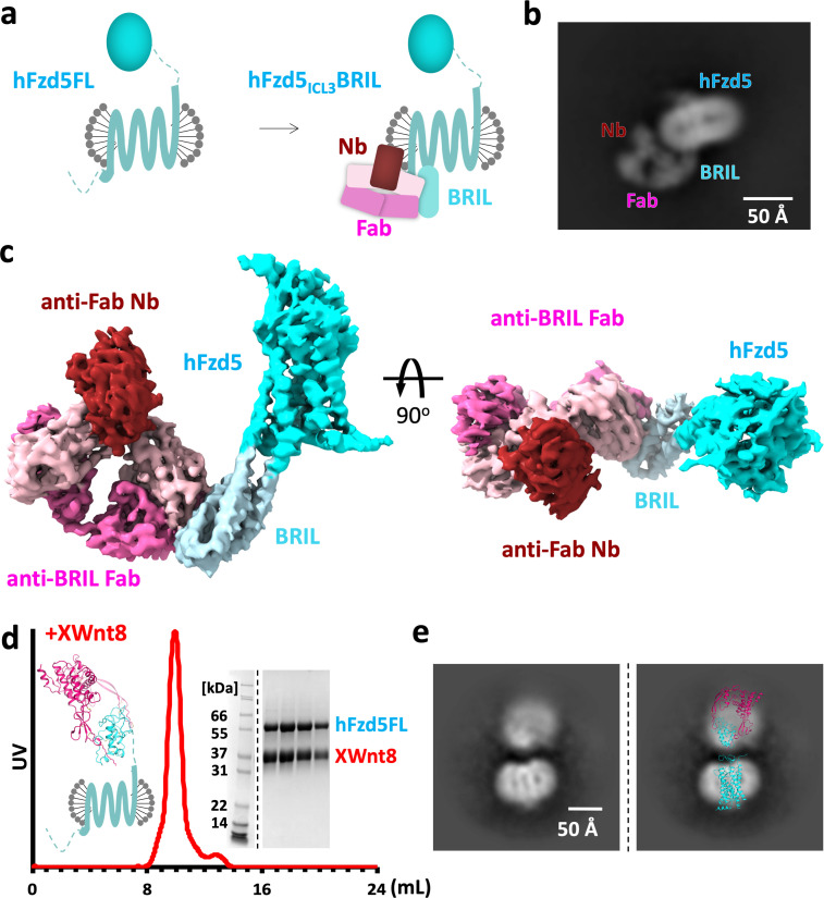
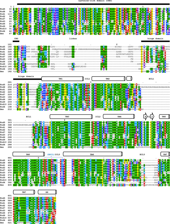
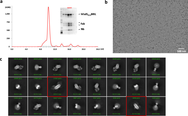
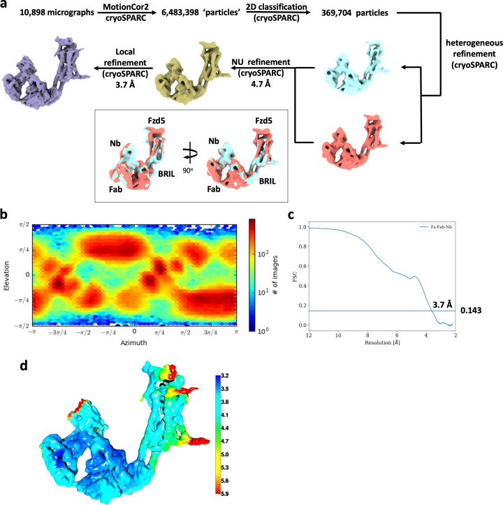
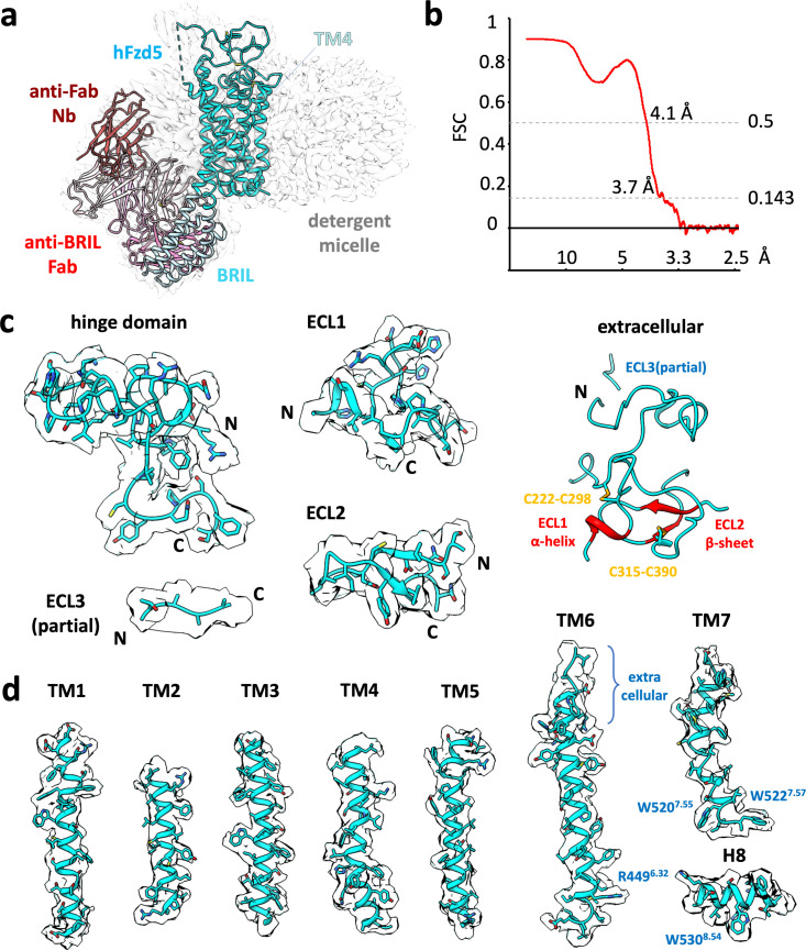
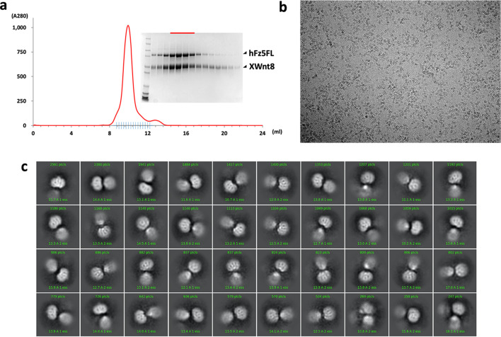
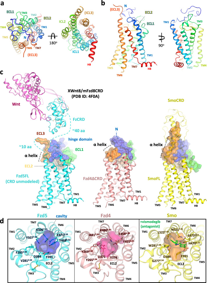
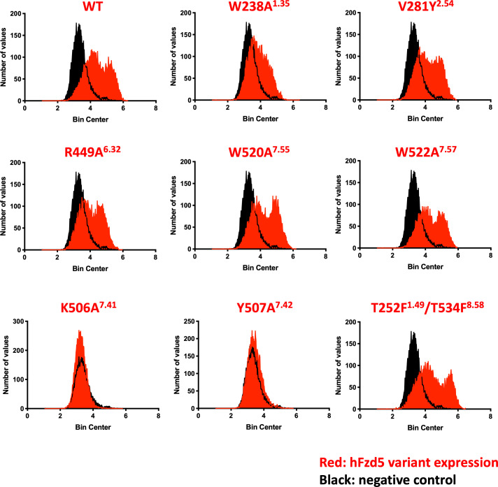
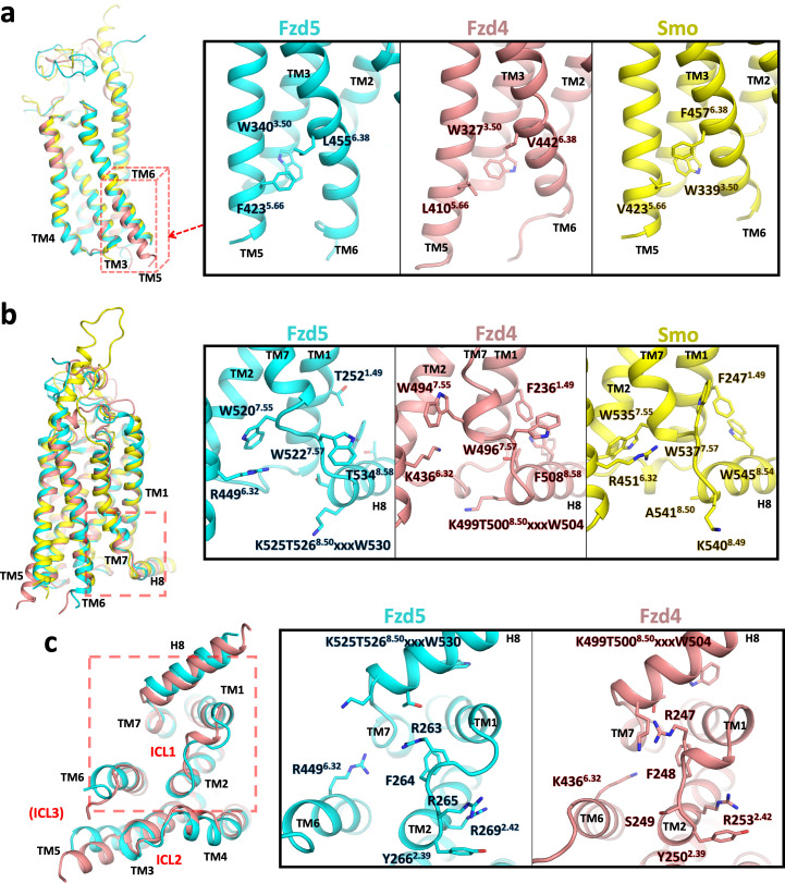
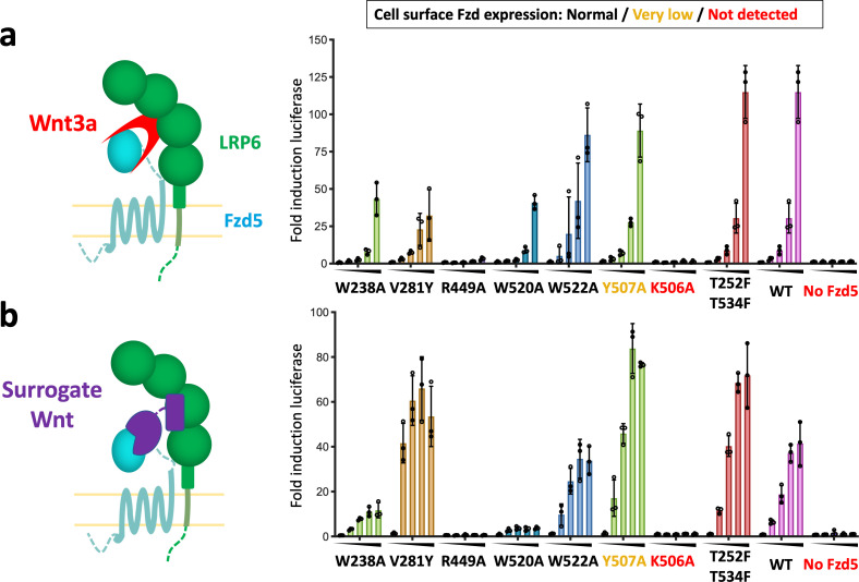
Frizzleds (Fzd) are the primary receptors for Wnt morphogens, which are essential regulators of stem cell biology, yet the structural basis of Wnt signaling through Fzd remains poorly understood. Here we report the structure of an unliganded human Fzd5 determined by single-particle cryo-EM at 3.7 Å resolution, with the aid of an antibody chaperone acting as a fiducial marker. We also analyzed the topology of low-resolution XWnt8/Fzd5 complex particles, which revealed extreme flexibility between the Wnt/Fzd-CRD and the Fzd-TM regions. Analysis of Wnt/β-catenin signaling in response to Wnt3a versus a 'surrogate agonist' that cross-links Fzd to LRP6, revealed identical structure-activity relationships. Thus, canonical Wnt/β-catenin signaling appears to be principally reliant on ligand-induced Fzd/LRP6 heterodimerization, versus the allosteric mechanisms seen in structurally analogous class A G protein-coupled receptors, and Smoothened. These findings deepen our mechanistic understanding of Wnt signal transduction, and have implications for harnessing Wnt agonism in regenerative medicine.

Keywords: Cryo-EM; Frizzled; Wnt; canonical Wnt/β-catenin signaling; human; molecular biophysics; structural biology; surrogate Wnt agonist.


Conflict of interest statement

NT, SM, DW, KJ, YM, JB, NA, AK, CG No competing interests declared, CJ, KG KCG and CYJ are founders of Surrozen Therapeutics.

Figures

Figure 1.
Figure 1.. Design scheme and cryo-EM analysis of hFzd5.
(a) Cartoon representation of the strategy for hFzd5 particle decoration by anti-BRIL Fab and anti-Fab Nb. These chaperones double hFzd5 molecular weight and render it asymmetric in detergent micelle, thus working as a fiducial maker for image alignment. (b) A selected 2D class average showing the side view of monomeric hFzd5ICL3BRIL/Fab/Nb. (c) Overall EM volume around the structural model of hFzd5 (cyan), BRIL and the linker between BRIL and Fzd5 (light blue), Fab heavy chain (pink), Fab light chain (light pink) and Nb (wine). (d) The size-exclusion chromatography profile, SDS-PAGE of peak fractions, and (e) the selected 2D class average of XWnt8/hFzd5FL. The full SDS-PAGE and 2D classes are shown in Figure 1—figure supplement 5. Models of XWnt8/mouse Fzd8CRD (mFzd8CRD) (PDB ID: 4F0A) and hFzd5 are overlaid on the blobs to show the relative size of densities.
Figure 1—figure supplement 1.
Figure 1—figure supplement 1.. Multiple sequence alignment of human class F GPCRs.
Sequence alignment of CRD and transmembrane regions of human class F GPCRs. The CRD region and individual transmembrane helix is labelled above the amino acid sequence. The alignments for CRD-hinge domain was generated using MAFFT (https://www.ebi.ac.uk/Tools/msa/mafft), and for TM1-H8 was taken from GPCR data base (www.gpcrdb.org). The alignment result was displayed using MView (www.ebi.ac.uk/Tools/msa/mview).
Figure 1—figure supplement 2.
Figure 1—figure supplement 2.. Purification, data collection and 2D classification of hFzd5ICL3BRIL/Fab/Nb.
(a) Representative size-exclusion chromatography profile of the hFzd5ICL3BRIL/Fab/Nb complex with the Coomassie stained SDS-PAGE around the peak fractions. SDS samples were prepared in reducing condition. The fractions used for EM are indicated by the red bar. (b) Representative raw micrograph of the hFzd5ICL3BRIL/Fab/Nb complex in detergent. (c) 2D class averages of selected particles. The dimer classes are marked by red boxes.
Figure 1—figure supplement 3.
Figure 1—figure supplement 3.. Cryo-EM data analysis of hFzd5ICL3BRIL/Fab/Nb.
(a) Cryo-EM data processing scheme. The resulting maps from the final heterogeneous refinement are superimposed for comparison (cyan and brick). (b) Orientation distribution of particles for the final 3D reconstruction. (c) The gold standard FSC curve for the final map. (d) Local resolution estimation of the final map colored in rainbow.
Figure 1—figure supplement 4.
Figure 1—figure supplement 4.. Cryo-EM map of representative built in model.
(a) Cryo-EM map (gray) and structural model of hFzd5ICL3BRIL in detergent complexed with anti-BRIL Fab and anti-Fab Nb. The model is colored as follow: the sequences from Fzd5 (cyan), BRIL and the linker between BRIL and Fzd5 (light blue), Fab heavy chain (pink), Fab light chain (light pink) and Nb (brown). (b) The map-model FCS curve generated by Phenix refinement (masked). Cryo-EM volumes (pale cyan) around the (c) extracellular regions and (d) individual transmembrane helices of hFzd5. Except for the whole extracellular region (c right) which only shows disulfide cysteine resides with red colored secondary structures assigned by DSSP, all sidechains are displayed regardless of their quality or model placement.
Figure 1—figure supplement 5.
Figure 1—figure supplement 5.. Purification, data collection and 2D class averages of the XWnt8/hFzd5FL complex.
(a) A size-exclusion chromatography profile of the XWnt8/hFzd5FL complex with the Coomassie stained SDS-PAGE. SDS samples were prepared in reducing condition. The peak fractions for the EM analysis are indicated by the red bar. (b) Representative raw micrograph of the XWnt8/hFzd5 complex. (c) 2D class averages of selected particles.
Figure 2.
Figure 2.. Overall structure of hFzd5 and structural comparison with hFzd4 and hSmo.
(a) Top (left) and bottom (right) views, and (b) side views of hFzd5 colored by rainbow with blue on the N-terminus and red on the C-terminus of the structural model. (c) Comparison of the extracellular regions between hFzd5, hFzd4 (pink, PDB ID: 6BD4) and hSmo (yellow, PDB ID: 5L7D). Transparent surfaces are overlaid on the cartoon models and colored separately by the hinge domain (blue), ECL1 (green), ELC2 (wheat) and ELC3 (orange). XWnt8/mFzd8CRD structure (PDB ID: 4F0A) is displayed and connected to the hFzd5 structure by dashed line, to show the size of the LRP6 binding module relative to 7TM. The α-helices at TM6-ECL3 are indicated with gray dashed lines. (d) Top views of hFzd5, hFzd4, and hSmo around the Smo-7TM ligand binding site with potential gatekeeping residues. The cavity volume calculated using the CavityPlus server (Xu et al., 2018) are displayed as transparent surface representations, with a focus on the constrictions made by bulky resides of Fzd at the upper-core site. The antagonist vismodegib (from PDB ID: 5L7I) is overlaid on the hSmo structure in its inactive and apo state (PDB ID: 5L7D).
Figure 2—figure supplement 1.
Figure 2—figure supplement 1.. Confirmation of hFzd5 receptor expression in HEK293T transfected cells.
Cell-surface hFzd5 variants were fluorescently labeled, and frequency distribution of FACS data are presented for each variant (red) with a negative control (black).
Figure 3.
Figure 3.. Conformational differences between hFzd5, hFzd4 and hSmo.
(a, b) Structural comparison of hFzd5, hFzd4 and hSmo (a) at the cytoplasmic end of TM5, and (b) the K/R6.32-W7.55 ionic lock with the aromatic network around H8. (c) Bottom views of hFzd5 and hFzd4 showing structural rearrangement of ICL1 and H8 important for Dvl binding. Each molecule is colored by cyan (hFzd5), pink (hFzd4, PDB ID: 6BD4) or yellow (Smo, PDB ID: 5L7D).
Figure 4.
Figure 4.. Structure-guided hFzd5 mutant signaling assays.
Schematic presentation of the ternary complex formation, and hFzd5-mediated signaling upon stimulation by (a) Wnt3a and (b) surrogate Wnt agonist. Non-lipidated, water-soluble surrogate Wnt would form the complex in a different geometry from natural Wnt ligands. Each series was stimulated (a) by Wnt3a at the concentrations of 0, 1.6, 3.1, 6.3 or 12.5 nM, or (b) by surrogate Wnt at the concentrations of 0, 0.25, 1, 4 or 16 nM, displayed from left (no agonist) to right (the highest concentration) and indicated by black triangles. The bars and error bars represent means and standard deviations, respectively, of data points from three technical replicates shown as circles.

References

    1. Adams PD, Afonine PV, Bunkóczi G, Chen VB, Davis IW, Echols N, Headd JJ, Hung LW, Kapral GJ, Grosse-Kunstleve RW, McCoy AJ, Moriarty NW, Oeffner R, Read RJ, Richardson DC, Richardson JS, Terwilliger TC, Zwart PH. PHENIX: a comprehensive Python-based system for macromolecular structure solution. Acta Crystallographica Section D Biological Crystallography. 2010;66:213–221. doi: 10.1107/S0907444909052925. - DOI - PMC - PubMed
    1. Anastas JN, Moon RT. WNT signalling pathways as therapeutic targets in Cancer. Nature Reviews Cancer. 2013;13:11–26. doi: 10.1038/nrc3419. - DOI - PubMed
    1. Audet M, White KL, Breton B, Zarzycka B, Han GW, Lu Y, Gati C, Batyuk A, Popov P, Velasquez J, Manahan D, Hu H, Weierstall U, Liu W, Shui W, Katritch V, Cherezov V, Hanson MA, Stevens RC. Crystal structure of misoprostol bound to the labor inducer prostaglandin E2 receptor. Nature Chemical Biology. 2019;15:11–17. doi: 10.1038/s41589-018-0160-y. - DOI - PMC - PubMed
    1. Bang I, Kim HR, Beaven AH, Kim J, Ko SB, Lee GR, Kan W, Lee H, Im W, Seok C, Chung KY, Choi HJ. Biophysical and functional characterization of norrin signaling through Frizzled4. PNAS. 2018;115:8787–8792. doi: 10.1073/pnas.1805901115. - DOI - PMC - PubMed
    1. Byrne EFX, Sircar R, Miller PS, Hedger G, Luchetti G, Nachtergaele S, Tully MD, Mydock-McGrane L, Covey DF, Rambo RP, Sansom MSP, Newstead S, Rohatgi R, Siebold C. Structural basis of smoothened regulation by its extracellular domains. Nature. 2016;535:517–522. doi: 10.1038/nature18934. - DOI - PMC - PubMed

Publication types