

Cardiac Involvement in Patients Recovered From COVID-2019 Identified Using Magnetic Resonance Imaging
- PMID: 32763118
- PMCID: PMC7214335
- DOI: 10.1016/j.jcmg.2020.05.004
Cardiac Involvement in Patients Recovered From COVID-2019 Identified Using Magnetic Resonance Imaging
Abstract
Objectives: This study evaluated cardiac involvement in patients recovered from coronavirus disease-2019 (COVID-19) using cardiac magnetic resonance (CMR).
Background: Myocardial injury caused by COVID-19 was previously reported in hospitalized patients. It is unknown if there is sustained cardiac involvement after patients' recovery from COVID-19.
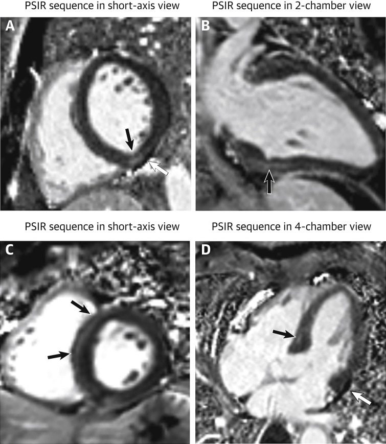
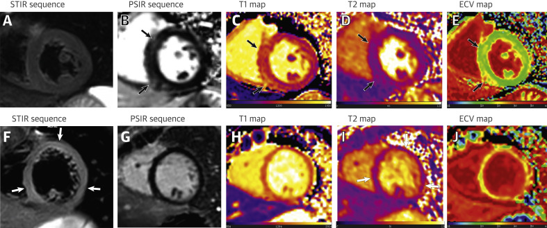
Methods: Twenty-six patients recovered from COVID-19 who reported cardiac symptoms and underwent CMR examinations were retrospectively included. CMR protocols consisted of conventional sequences (cine, T2-weighted imaging, and late gadolinium enhancement [LGE]) and quantitative mapping sequences (T1, T2, and extracellular volume [ECV] mapping). Edema ratio and LGE were assessed in post-COVID-19 patients. Cardiac function, native T1/T2, and ECV were quantitatively evaluated and compared with controls.
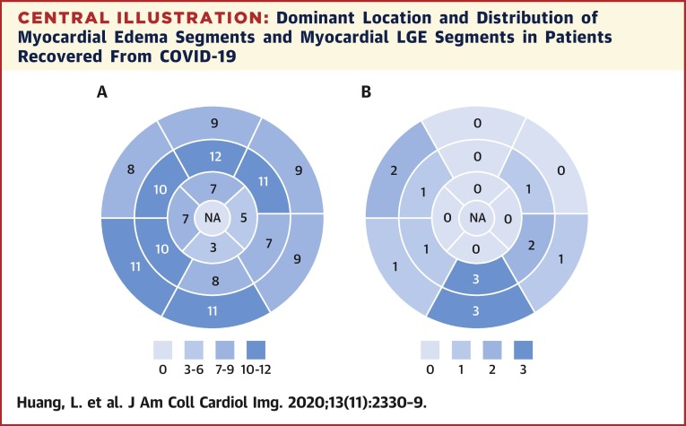
Results: Fifteen patients (58%) had abnormal CMR findings on conventional CMR sequences: myocardial edema was found in 14 (54%) patients and LGE was found in 8 (31%) patients. Decreased right ventricle functional parameters including ejection fraction, cardiac index, and stroke volume/body surface area were found in patients with positive conventional CMR findings. Using quantitative mapping, global native T1, T2, and ECV were all found to be significantly elevated in patients with positive conventional CMR findings, compared with patients without positive findings and controls (median [interquartile range]: native T1 1,271 ms [1,243 to 1,298 ms] vs. 1,237 ms [1,216 to 1,262 ms] vs. 1,224 ms [1,217 to 1,245 ms]; mean ± SD: T2 42.7 ± 3.1 ms vs. 38.1 ms ± 2.4 vs. 39.1 ms ± 3.1; median [interquartile range]: 28.2% [24.8% to 36.2%] vs. 24.8% [23.1% to 25.4%] vs. 23.7% [22.2% to 25.2%]; p = 0.002; p < 0.001, and p = 0.002, respectively).
Conclusions: Cardiac involvement was found in a proportion of patients recovered from COVID-19. CMR manifestation included myocardial edema, fibrosis, and impaired right ventricle function. Attention should be paid to the possible myocardial involvement in patients recovered from COVID-19 with cardiac symptoms.
Keywords: ACE2, angiotensin-converting enzyme 2; AHA, American Heart Association; BSA, body surface area; CI, cardiac index; CMR, cardiac magnetic resonance; CO, cardiac output; COVID-19, coronavirus disease-2019; ECV, extracellular volume; EDV, end-diastolic volume; EF, ejection fraction; ER, edema ratio; ESV, end-systolic volume; FA, flip angle; FOV, field of view; IQR, interquartile range; LGE, late gadolinium enhancement; LV, left ventricle; LVEF, left ventricular ejection fraction; PSIR, phase-sensitive inversion-recovery; RT-PCR, reverse transcription and polymerase chain reaction; RV, right ventricle; RVEF, right ventricular ejection fraction; SARS-CoV-2, severe acute respiratory syndrome-coronavirus-2; SI, signal intensity; SSFP, steady state free precession; STIR, short tau inversion recovery; SV, stroke volume; T2WI, T2-weighted imaging; TE, echo time; TR, repetition time; cardiac involvement; cardiac magnetic resonance imaging; coronavirus disease-2019; hs-cTnI, high-sensitive cardiac troponin I.
© 2020 by the American College of Cardiology Foundation. Published by Elsevier.
Conflict of interest statement
This work was supported in part by the National Natural Science Foundation of China (81471637 and 81873889), the National Mega Project on Major Infectious Disease Prevention (2017ZX10103005-007), and the National Key Research and Development Program of China (2018YFE0204500). All authors have reported that they have no relationships relevant to the contents of this paper to disclose.
Figures

Comment in
-
CMR in the Era of COVID-19: Evaluation of Myocarditis in the Subacute Phase.JACC Cardiovasc Imaging. 2020 Nov;13(11):2340-2342. doi: 10.1016/j.jcmg.2020.06.013. Epub 2020 Jul 3. JACC Cardiovasc Imaging. 2020. PMID: 32771570 Free PMC article.
-
Cardiac Involvement in the COVID-19 Pandemic: Hazy Lessons From Cardiac Imaging?JACC Cardiovasc Imaging. 2020 Nov;13(11):2480-2483. doi: 10.1016/j.jcmg.2020.10.001. Epub 2020 Oct 10. JACC Cardiovasc Imaging. 2020. PMID: 33153538 Free PMC article. No abstract available.
References
-
- World Health Organization WHO Director-General’s opening remarks at the media briefing on COVID-19 - 30 March 2020. World Health Organization. https://www.who.int/dg/speeches/detail/who-director-general-s-opening-re... Available at:
Publication types
MeSH terms
LinkOut - more resources
Full Text Sources
Medical
Research Materials
Miscellaneous
