. 2021 Jan;74(1):156-167.
doi: 10.1016/j.jhep.2020.07.041. Epub 2020 Aug 4.

Hepatocyte pyroptosis and release of inflammasome particles induce stellate cell activation and liver fibrosis

Affiliations

Hepatocyte pyroptosis and release of inflammasome particles induce stellate cell activation and liver fibrosis

Susanne Gaul et al. J Hepatol. 2021 Jan.

Abstract

Background & aims: Increased hepatocyte death contributes to the pathology of acute and chronic liver diseases. However, the role of hepatocyte pyroptosis and extracellular inflammasome release in liver disease is unknown.

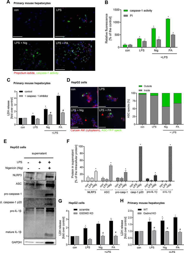
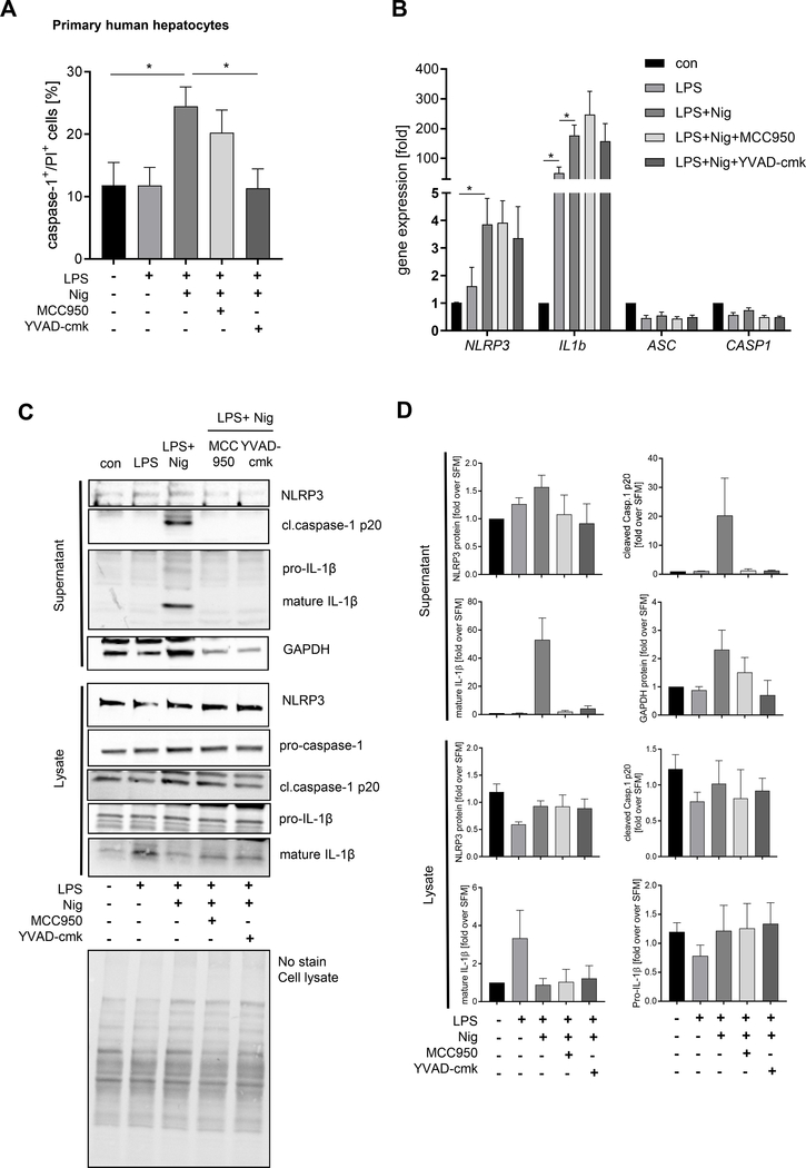
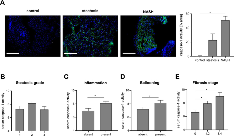
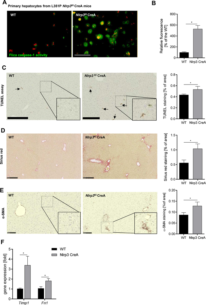
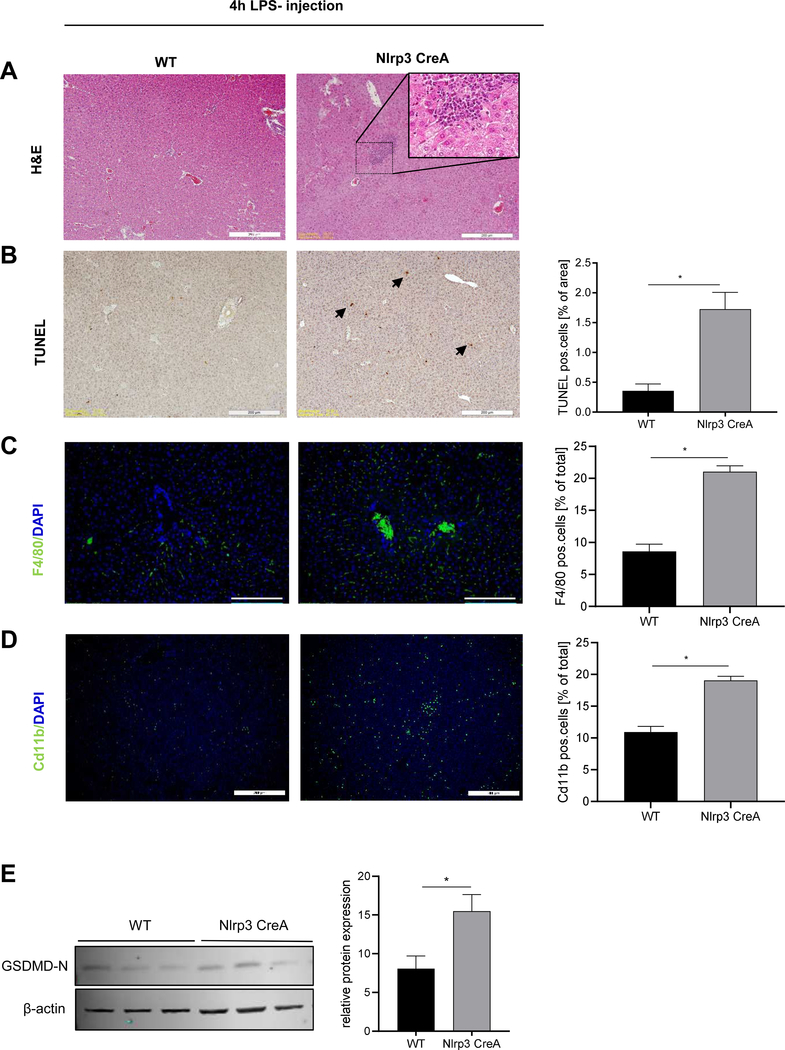
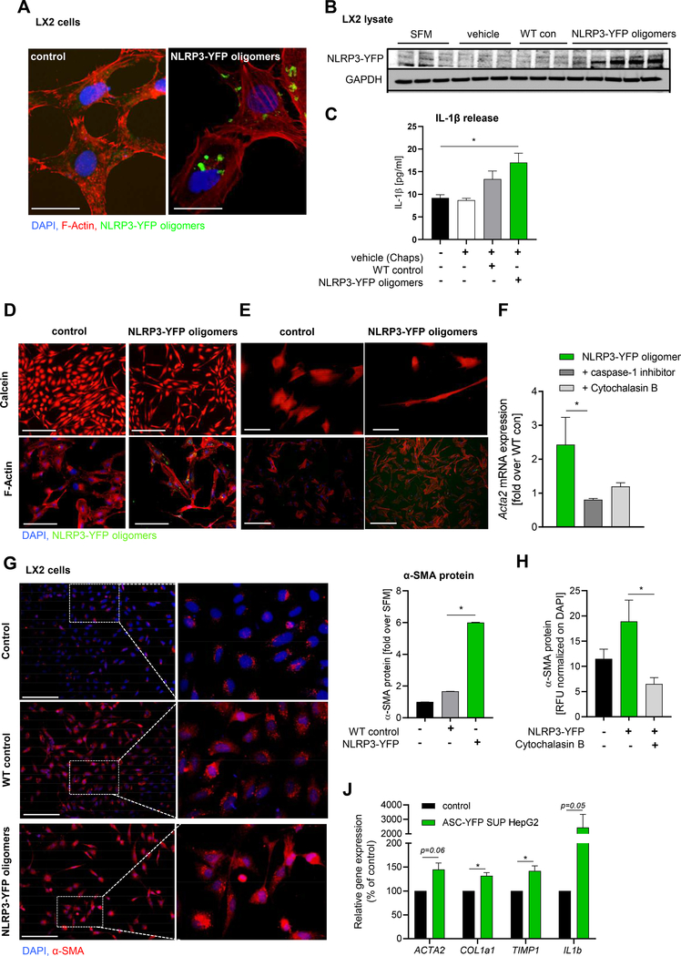
Methods: We used primary mouse and human hepatocytes, hepatocyte-specific leucine 351 to proline Nlrp3KICreA mice, and GsdmdKO mice to investigate pyroptotic cell death in hepatocytes and its impact on liver inflammation and damage. Extracellular NOD-, LRR-, and pyrin domain-containing protein 3 (NLRP3) inflammasomes were isolated from mutant NLRP3-YFP HEK cells and internalisation was studied in LX2 and primary human hepatic stellate cells. We also examined a cohort of 154 adult patients with biopsy-proven non-alcoholic fatty liver disease (Sir Charles Gairdner Hospital, Nedlands, Western Australia).

Results: We demonstrated that primary mouse and human hepatocytes can undergo pyroptosis upon NLRP3 inflammasome activation with subsequent release of NLRP3 inflammasome proteins that amplify and perpetuate inflammasome-driven fibrogenesis. Pyroptosis was inhibited by blocking caspase-1 and gasdermin D activation. The activated form of caspase-1 was detected in the livers and in serum from patients with non-alcoholic steatohepatitis and correlated with disease severity. Nlrp3KICreA mice showed spontaneous liver fibrosis under normal chow diet, and increased sensitivity to liver damage and inflammation after treatment with low dose lipopolysaccharide. Mechanistically, hepatic stellate cells engulfed extracellular NLRP3 inflammasome particles leading to increased IL-1β secretion and α-smooth muscle actin expression. This effect was abrogated when cells were pre-treated with the endocytosis inhibitor cytochalasin B.

Conclusions: These results identify hepatocyte pyroptosis and release of inflammasome components as a novel mechanism to propagate liver injury and liver fibrosis development.

Lay summary: Our findings identify a novel mechanism of inflammation in the liver. Experiments in cell cultures, mice, and human samples show that a specific form of cell death, called pyroptosis, leads to the release of complex inflammatory particles, the NLRP3 inflammasome, from inside hepatocytes into the extracellular space. From there they are taken up by other cells and thereby mediate inflammatory and pro-fibrogenic stress signals. The discovery of this mechanism may lead to novel treatments for chronic liver diseases in the future.

Keywords: ASC; Fibrosis; Hepatocytes; Inflammasome; Liver; NASH; NLRP3; Pyroptosis; Specks.


Conflict of interest statement

Conflict of interest The authors declare no conflicts of interest that pertain to this work. Please refer to the accompanying ICMJE disclosure forms for further details.

Figures

Figure 1.
Figure 1.. Hepatocytes undergo pyroptotic cell death following NLRP3 inflammasome activation
Representative immunofluorescence images (A) and quantification (B) showing pyroptosis induction by LPS + Nig and LPS + PA in primary mouse hepatocytes (*p< 0.05 compared to control, #p< 0.05 compared to LPS) (scale bar: 100 μm). (C) LDH release in presence of caspase-1 inhibitor (*p< 0.05 compared to control, #p< 0.05 compared to LPS + Nig and LPS + PA, respectively). (D) Representative images showing release of ASC specks from HepG2 cells transfected with a plasmid expressing ASC-YFP and treated with LPS + Nig and LPS + PA (scale bar: 10 0μm). Intra- an dextracellula rspeck swer equantified and compared to control. Western blot (E) and densitometric analysis (F) of cell-free supernatant of HepG2 cells after treatment with LPS and LPS + Nig for NLRP3 inflammasome components (NLRP3, ASC, pro-caspase1, pro-ILf) an dit scleave dmatur eform s(cleaved caspase-1 p20, mature IL-1ß 17kDa). Level of extracellular proteins were calculated as % of their intracellular protein amount (F) (*p< 0.05 vs. control). (G) LDH release of Gasdermin D (KD) silenced or scramble siRNA transfected HepG2 cells treated with LPS + Nig or PA. (H) LDH release frol primar ymurin ehepatocyte so fGsdmdK Ocompare dt oW Tmice treated with LPS + Nig or PA. Data was normalized to control serum-free media (*p< 0.05 vs. control, #p<0.05 vs. WT hepatocytes). Gaussian distributed data were analyzed using One-way analysis of variance and Bonferroni post-hoc test.
Figure 2.
Figure 2.. Primary human hepatocytes (PHH) undergo pyroptotic cell death and release activated inflammasome proteins and IL-1ß
PHH (n=3–4) were treated in starvation medium for NLRP3 inflammasome activation with LPS and Nig and in presence of inhibitors for NLRP3 and caspase-1. (A) Flow cytometric analysis of compensated Flica-caspase-1and PI double-positive cells (*p< 0.05). (B) mRMA expression of primary human hepatocytes (n=3) treated with LPS + Nig with or without NLRP3 and caspase-1 inhibitors normalized on housekeeping genes and referred to the control which was set as 1. Statistical significance was tested using One-way ANOVA and Bonferroni post-hoc test (*p< 0.05). Western blot analysis (C) and densitometric analysis (D) of concentrated supernatant and lysate of human hepatocytes (n=3) for NLRP3, cleaved caspase-1 p20, pro-caspase-1, mature IL-1ß 17kDa and pro-IL1ß normalized on whole-lane sample load. Data were referred to starvation media which was set at 1 (D).
Figure 3.
Figure 3.. Caspase-1 activity increases in liver and serum of NASH patients and correlates with liver injury severity
(A) Active caspase-1 levels were analyzed in frozen liver tissue sections (n 3 per group). Nuclei were stained with DAPI. Fluorescence intensity was analyzed using ImageJ and normalized on DAPI positive cells. Statistical significance was tested using One-way ANOVA and Bonferroni post-hoc test. (*p< 0.05 vs. control, scale bar: 100 μm). Level of serum caspase-1 activity in patients with present or absent (B) Steatosis, (C) Inflammation, (D) Ballooning and (E) Fibrosis stage. Two groups were analyzed by Studentś T-test (*p< 0.05 vs. control).
Figure 4.
Figure 4.. Hepatocyte-specific Nlrp3 mutant mice (L351P Nlrp3KICreA) showed increased hepatocyte caspase-1 activation and increased fibrogenesis
(A) Flica-caspase-1 activity (green) and PI (red) stained Nlrp3KICreA and WT primary hepatocytes (scale bar: 100 μm). (B) Quantification of caspase-1 positive cells (%) normalized on WT hepatocytes. (*p< 0.05 vs. WT hepatocytes). Immunohistological stainings and quantification of livers from WT and Nlrp3KICreA mice for (C) TUNEL positive cells (scale bar: 250 μm), (D) Sirius red collagen disposition (scale bar: 250 μm), and (E) α-SMA protein (scale bar: 100 μm). (F) mRNA expression of Timp1 and Fibronectin 1 (Fn1) in Nlrp3KICreA livers. Two groups were analyzed by Studentś T-test (*p< 0.05).
Figure 5.
Figure 5.. Hepatocyte-specific Nlrp3 mutant mice are more prone to LPS- induced liver inflammation and hepatocytes cell death
(A) H&E staining, (B) TUNEL positive cells (% of total area), (C) F4/80 positive cells and (D) Cd11b positive cells (% of total cells) in livers from WT and Nlrp3KICreA mice injected with LPS (scale bars: 200 μm). (E) Western blot and densitometric analysis of GasderminD N-terminal (31 kDa) protein in liver lysate from WT (n=3) and Nlrp3KICreA mice (n=3) injected with LPS. Data were normalized on β-actin. Two groups were analyzed by Studentś T-tes (*p< 0.05 vs. WT control).
Figure 6.
Figure 6.. Hepatic stellate cells internalize extracellular NLRP3/ASC inflammasome specks that leads to their activation
(A) Immunocytochemistry of LX2 cells incubated with extracellular NLRP3-YFP inflammasome particles and stained with F-Actin (red) and nuclei (DAPI, blue) (scale bar: 25 μm). (B) Western blot analysis and (C) IL-1ß ELISA of LX2 cells (n=3) treated with vehicle (n=3), WT control particles (WT con) (n=3) and NLRP3-YFP oligomeric particles (n=5). Primary antibody against YFP- tag and GAPDH was used. Data were compared using One-way analysis of variance and Bonferroni post-hoc test (*p< 0.05 compared to untreated control). Representative Calcein-stained and F-actin stained LX2 cells (n=4) (D) and primary human hepatic stellate cells (n=2) (E) stimulated with extracellular NLRP3-YFP oligomeric particles (green) for 24 h. Nuclei were stained with DAPI (blue) (scale bars: 100 μm). (F) mRNA expression of ACTA2 in NLRP3-YFP particles treated LX2 cells with or without caspase-1 inhibitor and endocytosis inhibitor Cytochalasin B. (G) Immunofluorescent α-smooth muscle actin (α-SMA) staining and quantification of LX2 cells stimulated with NLRP3-YFP oligomers or WT control particles for 24 h (scale bar: 100 μm). (H) Quantitated α-SMA immunofluorescent staining of LX2 cells treated with NLRP3-YFP oligomeric particles and Cytochalasin B for 24h. Relative fluorescence intensity (RFU) was normalized on total cells (DAPI). (J) mRNA expression of ACTA2, COL1a1, TIMP1 and IL1b in LX2 cells treated with supernatant of pyroptotic ASC-YFP transfected HepG2 cells compared to supernatant from untreated HepG2 cells. Two groups were analyzed by unpaired Studentś T-test (*p< 0.05).

References

    1. Luedde T, Kaplowitz N, Schwabe RF. Cell death and cell death responses in liver disease: mechanisms and clinical relevance. Gastroenterology 2014;147(4):765–783.e4. - PMC - PubMed
    1. Wree A, Mehal WZ, Feldstein AE. Targeting Cell Death and Sterile Inflammation Loop for the Treatment of Nonalcoholic Steatohepatitis. Seminars in liver disease 2016;36(1):27–36. - PMC - PubMed
    1. Broz P, Pelegrín P, Shao F. The gasdermins, a protein family executing cell death and inflammation. Nature reviews. Immunology 2020;20(3):143–57. - PubMed
    1. Strowig T, Henao-Mejia J, Elinav E, Flavell R. Inflammasomes in health and disease. Nature 2012;481(7381):278–86. - PubMed
    1. Broz P, Dixit VM. Inflammasomes: mechanism of assembly, regulation and signalling. Nature reviews. Immunology 2016;16(7):407–20. - PubMed

Publication types

MeSH terms