. 2020 Aug 5;21(16):5595.
doi: 10.3390/ijms21165595.

The Sphingosine Kinase 1 Inhibitor, PF543, Mitigates Pulmonary Fibrosis by Reducing Lung Epithelial Cell mtDNA Damage and Recruitment of Fibrogenic Monocytes

Affiliations

The Sphingosine Kinase 1 Inhibitor, PF543, Mitigates Pulmonary Fibrosis by Reducing Lung Epithelial Cell mtDNA Damage and Recruitment of Fibrogenic Monocytes

Paul Cheresh et al. Int J Mol Sci. .

Abstract

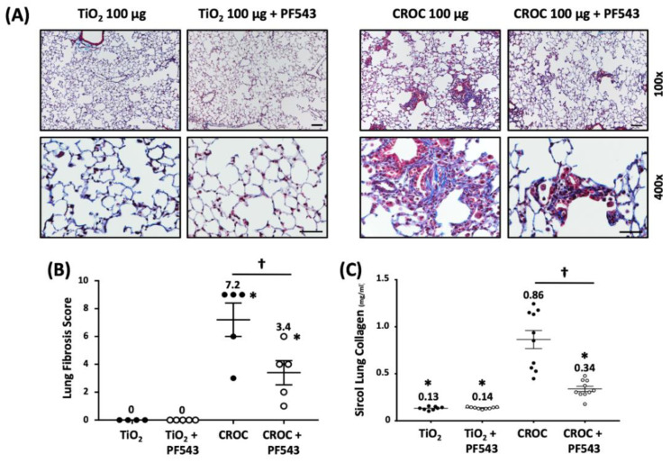
Idiopathic pulmonary fibrosis (IPF) is a chronic disease for which novel approaches are urgently required. We reported increased sphingosine kinase 1 (SPHK1) in IPF lungs and that SPHK1 inhibition using genetic and pharmacologic approaches reduces murine bleomycin-induced pulmonary fibrosis. We determined whether PF543, a specific SPHK1 inhibitor post bleomycin or asbestos challenge mitigates lung fibrosis by reducing mitochondrial (mt) DNA damage and pro-fibrotic monocyte recruitment-both are implicated in the pathobiology of pulmonary fibrosis. Bleomycin (1.5 U/kg), crocidolite asbestos (100 µg/50 µL) or controls was intratracheally instilled in Wild-Type (C57Bl6) mice. PF543 (1 mg/kg) or vehicle was intraperitoneally injected once every two days from day 7-21 following bleomycin and day 14-21 or day 30-60 following asbestos. PF543 reduced bleomycin- and asbestos-induced pulmonary fibrosis at both time points as well as lung expression of profibrotic markers, lung mtDNA damage, and fibrogenic monocyte recruitment. In contrast to human lung fibroblasts, asbestos augmented lung epithelial cell (MLE) mtDNA damage and PF543 was protective. Post-exposure PF543 mitigates pulmonary fibrosis in part by reducing lung epithelial cell mtDNA damage and monocyte recruitment. We reason that SPHK1 signaling may be an innovative therapeutic target for managing patients with IPF and other forms of lung fibrosis.

Keywords: SPHK1; alveolar epithelial cell; monocytes; mtDNA damage; oxidative stress; pulmonary fibrosis.


Conflict of interest statement

The authors declare no conflicts of interest.

Figures

Figure 1
Figure 1
Experimental Design. For the 21-d exposure, mice were treated with a single IT instillation of either bleomycin (1.5 U/kg), crocidolite asbestos (100 µg in 50 µL), or control (bleomycin control: PBS/DMSO−50 µL; asbestos control: TiO2 (100 µg/ 50 µL). On day 7 after IT instillation, the mice in each group were treated with interperitoneal (IP) instillation every other day with either PF543 (1 mg/kg) or saline until day 21. For the 60-d exposure, mice were treated with either asbestos or TiO2 as above and on day 30, the mice in each group were treated with either IP instillation of either PF543 (1 mg/kg) or saline every other day until day 60. At the end of treatment periods, the lungs were harvested for the various listed experimental endpoints.
Figure 2
Figure 2
Sphingosine Kinase 1 (SphK1) inhibition post bleomycin challenge mitigates pulmonary fibrosis in mice at 21 days. Fibrosis determined on day 21 in mouse lung. Hematoxylin and Eosin (A) and Trichrome staining (B) of lung specimens. Collagen levels by Sircol assay (C). All n = 5−8; * p < 0.05 vs. PBS+DMSO. Scale bars: 100× = 0.2 mm (Panel A), 50× = 1.0 mm (Panel B).
Figure 3
Figure 3
SphK1 Inhibition post challenge mitigates bronchoalveolar lavage (BAL) cells counts and protein markers of fibrosis in mice at 21 days. Fibrosis determined on day 21 (A) BAL cell counts. n = 6−8 (B) BAL protein and quantification of expression of fibronectin (FN), α-SMA, and collagen in lung tissues. n = 5−7 (C,D). All n = 4; * p < 0.05 vs. PBS+DMSO, PBS+DMSO+PF543, or PBS+ BLM+ PF543.
Figure 4
Figure 4
Sphk1 inhibition 7-days post-asbestos challenge attenuates pulmonary fibrosis in mice at 21 days. Fibrosis determined on day 21 in mouse lung. Trichrome staining (A) and fibrosis score (B) of lung specimens. Collagen levels by Sircol assay (C). n = 4−11; * p < 0.05 vs. Ti or Ti+PF543; p < 0.05 vs. Crocidolite asbestos (CROC). Scale bars: 100× = 0.1mm; 400× = 0.05 mm.
Figure 5
Figure 5
PF543 given at 30-days post-asbestos challenge attenuates pulmonary fibrosis in mice at 60 days. Fibrosis determined on day 60 in mouse lung. Trichrome staining (A) and fibrosis score (B) of lung specimens. Collagen levels by Sircol assay (C). n = 4−10; * p < 0.05 vs. Ti or Ti+PF543; p < 0.05 vs. CROC. Scale bars: 100× = 0.1 mm; 400× = 0.05 mm.
Figure 6
Figure 6
PF543 attenuates plasma S1P, lung mtDNA damage in asbestos-exposed mice at 21 d, and asbestos-induced mtDNA damage in Alveolar epithelial cell line MLE 12 (AEC-MLE-12) cells but not Human lung fibroblasts (HLF). Baseline plasma S1P levels (A) mtDNA damage in mouse lungs (B), and DNA damage in the nucleus and mitochondria of MLE-12 cells (C) and HLF (D). n = 3−5 * p < 0.05 vs. TiO2, p < 0.05 vs. CROC; and HLF (D), n = 3−5, * p < 0.05 vs. TiO2, p < 0.05 vs. CROC.
Figure 7
Figure 7
PF543 reduces the number of newly-made “fibrogenic” Mo AMs in mouse lungs exposed to crocidolite asbestos, 60-day post challenge in lungs of CXCR3 ER zsGREEN mice. Flow gating strategy (A). Number of new Mo-AMs recruited to lung (B). n = 4−8, * p < 0.05 vs. CROC alone.

References

    1. Martinez F.J., Collard H.R., Pardo A., Raghu G., Richeldi L., Selman M., Swigris J.J., Taniguchi H., Wells A.U. Idiopathic pulmonary fibrosis. Nat. Rev. Dis. Primers. 2017;3:17074. doi: 10.1038/nrdp.2017.74. - DOI - PubMed
    1. Olson A.L., Swigris J.J., Lezotte D.C., Norris J.M., Wilson C.G., Brown K.K. Mortality from pulmonary fibrosis increased in the United States from 1992 to 2003. Am. J. Respir. Crit. Care Med. 2007;176:277–284. doi: 10.1164/rccm.200701-044OC. - DOI - PubMed
    1. Huang L.S., Natarajan V. Sphingolipids in pulmonary fibrosis. Adv. Biol. Regul. 2015;57:55–63. doi: 10.1016/j.jbior.2014.09.008. - DOI - PMC - PubMed
    1. Shea B.S., Tager A.M. Role of the lysophospholipid mediators lysophosphatidic acid and sphingosine 1-phosphate in lung fibrosis. Proc. Am. Thorac. Soc. 2012;9:102–110. doi: 10.1513/pats.201201-005AW. - DOI - PMC - PubMed
    1. Pyne N.J., Dubois G., Pyne S. Role of sphingosine 1-phosphate and lysophosphatidic acid in fibrosis. Biochim. Biophys. Acta. 2013;1831:228–238. doi: 10.1016/j.bbalip.2012.07.003. - DOI - PubMed

MeSH terms

LinkOut - more resources