Review
. 2020 Aug 5;12(8):2179.
doi: 10.3390/cancers12082179.

The Biology of Exosomes in Breast Cancer Progression: Dissemination, Immune Evasion and Metastatic Colonization

Affiliations
Review

The Biology of Exosomes in Breast Cancer Progression: Dissemination, Immune Evasion and Metastatic Colonization

Cinzia Giordano et al. Cancers (Basel). .

Abstract

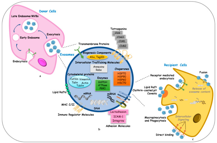
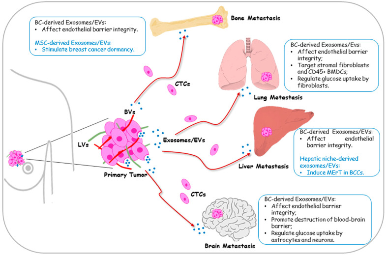
In recent decades, the study of exosome biology has gained growing interest, representing an active area of cancer research with many potential clinical applications. Exosomes are small lipid bilayer particles released by cells with pleiotropic functions that have been reported to regulate the complex intracellular pathway involved in all steps of breast cancer development-from initiation to progression toward a metastatic dissemination. Particularly, the role of these microvesicles has been explored in metastasis, which represents the leading cause of breast cancer morbidity and mortality worldwide. Reports highlight that the plasticity of breast cancer cells, fundamental for the establishment of distant metastasis, may be in part attributed to exosome-carried signals shared between adjacent cells and long-distance cells in the body. In the present review, we will discuss the functions of exosomes in the metastatic breast cancer process and secondary site outgrowth. The possibility to decode the exosome functions in advanced diseases may offer new opportunities for early detection, molecular targeted therapies and exosome-based therapeutics in breast cancer.

Keywords: EMT; breast cancer; exosomes; extracellular vesicles; metastasis.


Conflict of interest statement

The authors declare no conflict of interest.

Figures

Figure 1
Figure 1
Biogenesis, structure and uptake of exosomes. (a) Exosome biogenesis starts with membrane endocytosis resulting in the formation of early endosomes that subsequently mature into late endosomes or Multi Vesicular Bodies (MVBs). MVBs can be transported to the plasma membrane through the cytoskeletal network and exocytosis occurs, allowing the release of exosomes into the extracellular space. (b) Exosomes contain different families of proteins categorized according to their functions, lipids, metabolites and nucleic acids (DNA, RNA, miRNA and other non-coding RNAs). (c) The entry of exosomes in recipient cells can be mediated by several mechanisms: direct fusion of exosomes with the plasma membrane allowing the deposit of their content into the cytoplasm; receptor-mediated endocytosis, lipid rafts, clathrin-coated pit and caveolae, macropinicytosis, phagocytosis, that allow the entry of intact exosomes in the cells; direct binding of exosomes to specific receptors that induce intracellular signalings into recipient cells. HSC: heat shock cognate; HSP: heat shock protein; ICAM-1: intercellular adhesion molecule 1; MHC: major histocompatibility complex; PGK1: phosphoglycerate kinase 1; Rab: Ras-related proteins in brain; Tsg101: Tumor susceptibility gene 101.
Figure 2
Figure 2
Schematic representation of the effects of exosome/extracellular vesicles (EVs) released by tumor cells and stromal cells in the establishment of metastatic outgrowth in breast cancer. Primary breast cancer-derived exosomes drive organotropic metastasis of circulating tumor cells (CTCs) enabling their survival into blood (BVs) or lymphatic vessels (LVs) and establishing the permissive premetastatic niche at the distant sites: bone, lung, liver, and brain. The main mechanisms involved are summarized in the boxes near each metastatic site.

References

    1. Bray F., Ferlay J., Soerjomataram I., Siegel R.L., Torre L.A., Jemal A. Global cancer statistics 2018: GLOBOCAN estimates of incidence and mortality worldwide for 36 cancers in 185 countries. CA A Cancer J. Clin. 2018;68:394–424. doi: 10.3322/caac.21492. - DOI - PubMed
    1. Cardoso F., Costa A., Norton L., Cameron D., Cufer T., Fallowfield L., Francis P.A., Gligorov J., Kyriakides S., Lin N., et al. 1st International consensus guidelines for advanced breast cancer (ABC 1) Breast. 2012;21:242–252. doi: 10.1016/j.breast.2012.03.003. - DOI - PubMed
    1. Mariotto A.B., Etzioni R., Hurlbert M., Penberthy L., Mayer M. Estimation of the number of women living with metastatic breast cancer in the united states. Cancer Epidemiol. Biomark. Prev. 2017;26:809–815. doi: 10.1158/1055-9965.EPI-16-0889. - DOI - PMC - PubMed
    1. Harbeck N., Gnant M. Breast cancer. Lancet. 2017;389:1134–1150. doi: 10.1016/S0140-6736(16)31891-8. - DOI - PubMed
    1. Raposo G., Stoorvogel W. Extracellular vesicles: Exosomes, microvesicles, and friends. J. Cell Biol. 2013;200:373–383. doi: 10.1083/jcb.201211138. - DOI - PMC - PubMed

LinkOut - more resources