. 2020 Jul 17:12:5893-5902.
doi: 10.2147/CMAR.S257299. eCollection 2020.

Prognostic Significance of PD-L1 Expression and Its Tumor-Intrinsic Functions in Hypopharyngeal Squamous Cell Carcinoma

Affiliations

Prognostic Significance of PD-L1 Expression and Its Tumor-Intrinsic Functions in Hypopharyngeal Squamous Cell Carcinoma

Peng Cui et al. Cancer Manag Res. .

Abstract

Purpose: The expression of programmed death-ligand 1 (PD-L1) is common in various solid human cancers and it is an important therapeutic target. However, the expression pattern, clinical significance and potential mechanism of PD-L1 in hypopharyngeal squamous cell carcinoma (HSCC) are still lacking.

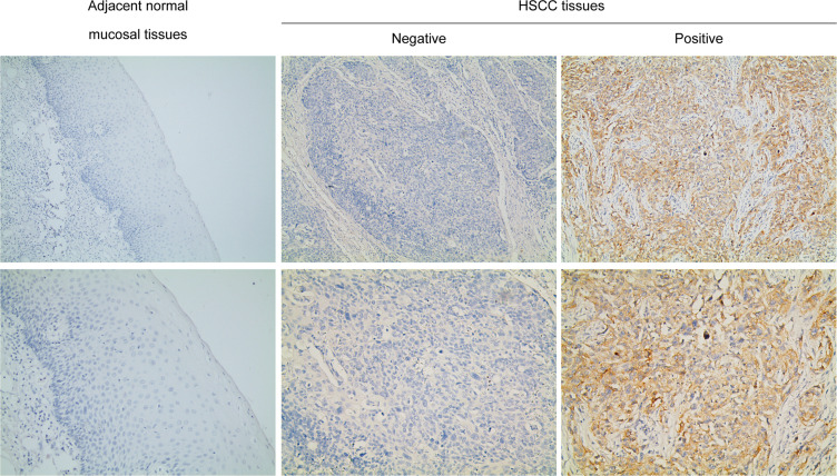
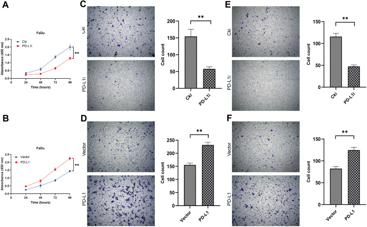
Methods: PD-L1 expression in HSCC tumor tissues and paired adjacent hypopharyngeal mucosal tissues was detected using immunohistochemistry assay, and the clinical significance of PD-L1 in HSCC was characterized. In vitro assays including cell viability assays, migration assays, invasion assays as well as Western blot assays were performed to illuminate the biological functions and underlying molecular mechanisms of PD-L1 in HSCC development.

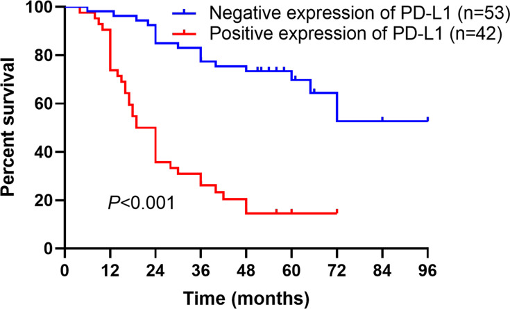
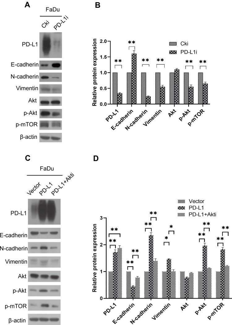
Results: PD-L1 expression was detected in HSCC samples but we found no positive expression in matched normal hypopharyngeal mucosal tissues. The levels of PD-L1 expression were significantly correlated with advanced clinical progression and poor patient survival. Multivariable analysis of Cox model showed that PD-L1 expression was an independent predictor for the prognosis of HSCC patients. Functional experiments showed that the ectopic expression of PD-L1 markedly influenced the proliferation, migration and invasion of FaDu cells in vitro. Mechanistically, investigations demonstrated that PD-L1 could promote the epithelial-mesenchymal transition of FaDu cells. Meanwhile, PD-L1 knockdown inhibited, while PD-L1 overexpression activated the Akt-mTOR signaling pathway in FaDu cells. The EMT induced by PD-L1 overexpression could be reversed by the Akt inhibitor.

Conclusion: In summary, the expression of PD-L1 can act as a significant biomarker for the adverse clinicopathological features and poor prognosis of patients with HSCC. PD-L1 can promote the proliferation, migration and invasion of FaDu cells and consequently enhance the aggressiveness. Moreover, PD-L1 induces EMT through AKT-mTOR signaling pathway. These suggest that PD-L1 has important tumor-intrinsic functions independent of its immunopathogenic effects.

Keywords: PD-L1; epithelial–mesenchymal transition; hypopharyngeal squamous cell carcinoma; prognosis.


Conflict of interest statement

The authors declare that there is no conflict of interest in this work.

Figures

Figure 1
Figure 1
PD-L1 expression in adjacent normal mucosal tissues and HSCC tissues. Representative immunohistochemical staining examples of PD-L1 protein expression are shown. In the current study there was no positive expression of PD-L1 in the mucosal tissues. Magnification, ×100 and ×200. Abbreviations: PD-L1, programmed death-ligand 1; HSCC, hypopharyngeal squamous cell carcinoma.
Figure 2
Figure 2
Kaplan-Meier analysis for overall survival of HSCC patients. Kaplan-Meier analysis of overall survival stratified by positive versus negative expression of PD-L1 in 95 HSCC patients (P<0.001). Log rank test was used for analyzing the differences. Abbreviations: HSCC, hypopharyngeal squamous cell carcinoma; PD-L1, programmed death-ligand 1.
Figure 3
Figure 3
PD-L1 promoted FaDu cells proliferation, migration and invasion in vitro. (A and B) Depletion of PD-L1 inhibited FaDu cells proliferation, whereas overexpression of PD-L1 stimulated FaDu cells proliferation as determined by CCK-8 assay. The data were presented as means ± standard deviation from three independent experiments. ** P<0.01. (C and D) The transwell migration assays showed that depletion of PD-L1 obviously inhibited the migration of FaDu cells. Conversely, overexpression of PD-L1 promoted the migration of FaDu cells. ** P<0.01. (E and F) The transwell invasion assays showed that depletion of PD-L1 obviously inhibited the invasion of FaDu cells. Conversely, overexpression of PD-L1 promoted the invasion of FaDu cells. The data were presented as means ± standard deviation from three independent experiments. **P<0.01. Abbreviations: PD-L1, programmed death-ligand 1; CCK-8, Cell Counting Kit-8 assay; Cki, control siRNA; siRNA, small interfering RNA.
Figure 4
Figure 4
PD-L1 promoted EMT through the Akt-mTOR signaling pathway. (A) FaDu cells were transfected with PD-L1 siRNA or control siRNA, expression of PD-L1, E-cadherin, N-cadherin, Vimentin, Akt, p-Akt and p-mTOR was analyzed by Western blot. β-actin was used as an inner control. (B) Bands of Western blot were analyzed by Image J software. Results were obtained from the ratio of target band to β-actin. The data are presented as means ± standard deviation from three independent experiments. **P<0.01. (C) PD-L1-overexpressed or control FaDu cells were treated with Akt-inhibitor. The indicated protein levels were assayed by Western blot. β-actin was used as an inner control. (D) Bands of Western blot were analyzed by Image J software. Results were obtained from the ratio of target band to β-actin. The data are presented as means ± standard deviation from three independent experiments. *P<0.05, **P<0.01. Abbreviations: PD-L1, programmed death-ligand 1; EMT, epithelial–mesenchymal transition; siRNA, small interfering RNA; Cki, control siRNA.

References

    1. Cooper JS, Porter K, Mallin K, et al. National cancer database report on cancer of the head and neck: 10-year update. Head Neck. 2009;31(6):748–758. doi:10.1002/hed.21022 - DOI - PubMed
    1. Hashibe M, Brennan P, Benhamou S, et al. Alcohol drinking in never users of tobacco, cigarette smoking in never drinkers, and the risk of head and neck cancer: pooled analysis in the International Head and Neck Cancer Epidemiology Consortium. J Natl Cancer Inst. 2007;99(10):777–789. doi:10.1093/jnci/djk179 - DOI - PubMed
    1. Carvalho AL, Nishimoto IN, Califano JA, Kowalski LP. Trends in incidence and prognosis for head and neck cancer in the United States: a site-specific analysis of the SEER database. Int J Cancer. 2005;114(5):806–816. doi:10.1002/ijc.20740 - DOI - PubMed
    1. Denaro N, Russi EG, Adamo V, Merlano MC. State-of-the-art and emerging treatment options in the management of head and neck cancer: news from 2013. Oncology. 2014;86(4):212–229. doi:10.1159/000357712 - DOI - PubMed
    1. Zhang Y, Wang B, Chen X, Li W, Dong P. AGO2 involves the malignant phenotypes and FAK/PI3K/AKT signaling pathway in hypopharyngeal-derived FaDu cells. Oncotarget. 2017;8(33):54735–54746. doi:10.18632/oncotarget.18047 - DOI - PMC - PubMed