

Optical Coherence Tomography Angiography in Neurodegenerative Diseases: A Review
- PMID: 32765149
- PMCID: PMC7368556
- DOI: 10.2147/EB.S193026
Optical Coherence Tomography Angiography in Neurodegenerative Diseases: A Review
Abstract
Background: Optical coherence tomography angiography (OCT-A) has emerged as a novel, fast, safe and non-invasive imaging technique of analyzing the retinal and choroidal microvasculature in vivo. OCT-A captures multiple sequential B-scans performed repeatedly over a specific retinal area at high speed, thus enabling the composition of a vascular map with areas of contrast change (high flow zones) and areas of steady contrast (slow or no flow zones). It therefore provides unique insight into the exact retinal or choroidal layer and location at which abnormal blood flow develops. OCTA has evolved into a useful tool for understanding a number of retinal pathologies such as diabetic retinopathy, age-related macular degeneration, central serous chorioretinopathy, vascular occlusions, macular telangiectasia and choroidal neovascular membranes of other causes. OCT-A technology is also increasingly being used in the evaluation of optic disc perfusion and has been suggested as a valuable tool in the early detection of glaucomatous damage and monitoring progression.
Objective: To review the existing literature on the applications of optical coherence tomography angiography in neurodegenerative diseases.
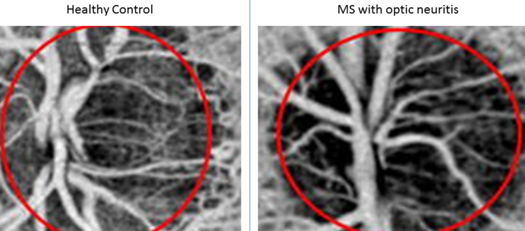
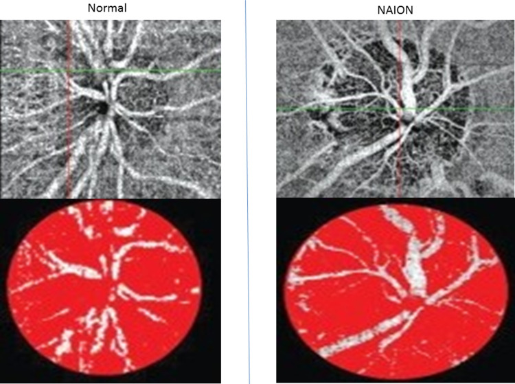
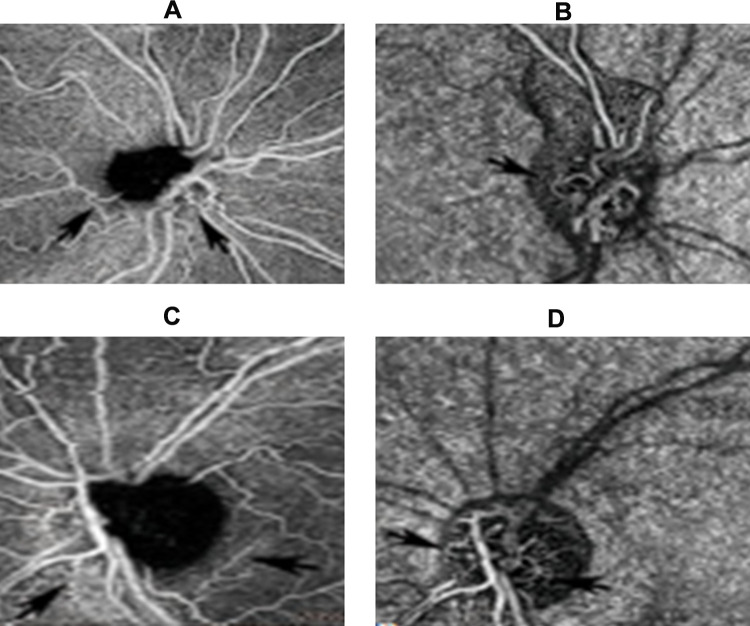
Summary: A meticulous literature was performed until the present day. Google Scholar, PubMed, Mendeley search engines were used for this purpose. We used 123 published manuscripts as our references. OCT-A has been utilized so far to describe abnormalities in multiple sclerosis (MS), Alzheimer's disease, arteritic and non-arteritic optic neuropathy (AION and NAION), Leber's hereditary optic neuropathy (LHON) papilloedema, Parkinson's disease, Huntington's disease, amyotrophic lateral sclerosis (ALS), Wolfram syndrome, migraines, lesions of the visual pathway and cerebral autosomal dominant arteriopathy with subcortical infarcts and leukoencephalopathy (CADASIL). It appears that OCT-A findings correlate quite well with the severity of the aforementioned diseases. However, OCT-A has its own limitations, namely its lack of wide-field view of the peripheral retina and the inaccurate interpretation due to motion artifacts in uncooperative groups of patients (e.g. children). Larger prospective longitudinal studies will need to be conducted in order to eliminate the aforementioned limitations.
Keywords: neuro-ophthalmology; neurodegenerative diseases; optical coherence tomography angiography.
© 2020 Tsokolas et al.
Conflict of interest statement
The authors report no conflicts of interest in this work. The authors alone are responsible for the content and writing of the paper.
Figures
References
Publication types
LinkOut - more resources
Full Text Sources
Other Literature Sources
Miscellaneous
