. 2020 Jul 10:11:1630.
doi: 10.3389/fmicb.2020.01630. eCollection 2020.

Heat Shock Protein Member 8 Is an Attachment Factor for Infectious Bronchitis Virus

Affiliations

Heat Shock Protein Member 8 Is an Attachment Factor for Infectious Bronchitis Virus

Pengpeng Zhu et al. Front Microbiol. .

Abstract

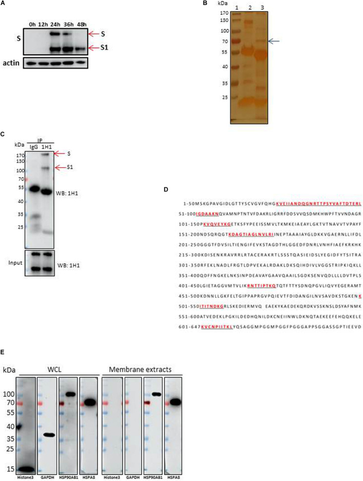
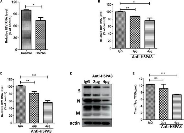
Although infectious bronchitis virus (IBV) is the first coronavirus identified, little is known about which membrane protein of host cells could interact with IBV spike protein and facilitate the infection by the virus. In this study, by using a monoclonal antibody to the S1 protein of IBV M41 strain, we found that heat shock protein member 8 (HSPA8) could interact with spike protein of IBV. HSPA8 was found to be present on the cell membrane and chicken tissues, with highest expression level in the kidney. Results of co-IP and GST-pull-down assays indicated that the receptor binding domain (RBD) of IBV M41 could interact with HSPA8. The results of binding blocking assay and infection inhibition assay showed that recombinant protein HSPA8 and antibody to HSPA8 could inhibit IBV M41 infection of chicken embryonic kidney (CEK) cells. Further, we found that HSPA8 interacted with the N-terminal 19-272 amino acids of S1 of IBV Beaudette, H120 and QX strains and HSPA8 from human and pig also interacted with IBV M41-RBD. Finally the results of binding blocking assay and infection inhibition assay showed that recombinant HSPA8 protein and antibody to HSPA8 could inhibit IBV Beaudette strain infection of Vero cells that were treated with heparanase to remove heparan sulfate from the cell surface. Taken together, our results indicate that HSPA8 is a novel host factor involved in IBV infection.

Keywords: HSPA8; attachment factor; infectious bronchitis virus; receptor binding domain; virus entry.


Figures

FIGURE 1
FIGURE 1
Identification of HSPA8 as a target membrane molecule for IBV Spike protein. (A) Determination of viral protein expression level of IBV M41 strain in CEK cells. CEK cells were infected with IBV M41 (TCID50 = 106.5/ml, 50 μl/well) in 6-well plates. Infected cells were harvested at 0, 12, 24, 36, and 48 h post-infection. The whole cell lysates were subjected to SDS-PAGE and Western blot analysis with mAb 1H1 to spike protein. The expression of cellular actin protein was used as control. (B) Silver staining of membrane proteins of CEK cells after immunoprecipitation assay. Membrane protein extracts of IBV M41 infected CEK cells were immunoprecipitated with mAb 1H1 (lane 3) to IBV M41 spike protein, and mouse IgG (lane 2). Lane 1, protein marker. Arrow indicated the protein band at 70kDa specifically precipitated by mAb 1H1. (C) Western blot analysis of membrane proteins of CEK cells after immunoprecipitation assay. Sample orders were same as shown in (B). (D) Amino acid sequence of HSPA8. The gel fragment indicated with the arrowhead in (B) was analyzed by LC-MS/MS. The resulting peptide sequences of HSPA8 were underlined and marked as red color. Full length HSPA8 sequence was searched against chicken protein database in Uniprot. (E) Western blot analysis of HSPA8 present in the membrane protein extracted from CEK cells. Membrane protein fraction of CEK cells and whole cell lysates of CEK cells were immunoblotted with antibodies against the cytoplasmic protein marker (GAPDH), nuclear protein marker (Histone3), membrane protein marker (HSP90AB1) and HSPA8. WCL, whole cell lysates.
FIGURE 2
FIGURE 2
Detection of expression of HSPA8 on cell surface and chicken tissues. (A,C,E) Identification of HSPA8 expression on surface of 293T cells (A), CEK cells (C) and Vero cells (E) by flow cytometry analysis with antibody against HSPA8. Total 6.5 × 10⁴ of 293T cells, CEK cells and Vero cells were used for each analysis. The blue lines mean without primary antibody and the red lines indicated the reaction with HSPA8 antibody. (B,D,F) Western blot analysis of the membrane protein extracts of 293T cells (B), CEK cells (D) and Vero cells (F) with antibody against HSPA8. (G) Detection of HSPA8 expression level in 1-day-old chicken tissues by RT-qPCR. Tissue samples collected from three chickens were used for the analysis of HSPA8 expression level. **P < 0.01; ***P < 0.001.
FIGURE 3
FIGURE 3
Interaction of HSPA8 with RBD of IBV spike protein. (A) Detection of the interaction between HSPA8 and IBV M41 spike protein in M41-infected CEK cells by immunoprecipitation assay using mAb 1H1 to IBV S and mouse IgG. IBV S1 and HSPA8 in precipitates were detected by immunoblots with mAbs to IBV S1 and HSPA8. (B) Detection of the interaction between HSPA8 and IBV M41-RBD by co-IP assay using HSPA8 as bait protein. 293T cells were cotransfected with indicated plasmids and then flag-tagged HSPA8 was immunoprecipitated with anti-flag antibody. The precipitated proteins were detected by Western blotting using anti-flag and anti-GFP antibodies. (C) Detection of the interaction between HSPA8 and IBV M41-RBD by reciprocal co-IP assay using M41-RBD as bait protein. 293T cells were cotransfected with indicated plasmids and then GFP-tagged M41-RBD was immunoprecipitated with anti-GFP antibody. The precipitated proteins were detected by Western blotting using anti-flag and anti-GFP antibodies. (D,E) Identification of the direct interaction between HSPA8 and IBV M41-RBD by GST-pull-down assay. In (D), GST and GST-M41-RBD recombinant proteins were separately bound to glutathione-sepharose beads and then incubated with His-HSPA8 recombinant protein. The protein complexes were detected by Western blot analysis with antibodies against GST and 6xHis. In (E), The lysates of 293T cells transfected with EGFP-C3-M41-RBD vector were incubated with GST or GST-HSPA8 protein which was separately bound to glutathione-sepharose beads. Subsequently, the protein complexes were detected by Western blot analysis with antibodies against GST and GFP.
FIGURE 4
FIGURE 4
Inhibition of IBV M41 binding to CEK cells by binding blocking assay and infection inhibition assay. (A) Recombinant HSPA8 blocked the binding of IBV M41 to CEK cells. IBV M41 (TCID50 = 106.5/ml, 200 μl) was incubated with recombinant protein GST-HSPA8 (100 μg) and control recombinant protein GST (100 μg) at 37°C for 1 h. CEK cells cultured in 6-well plates was incubated with GST-HSPA8-treated virus and GST-treated virus at 4°C for 1 h. After 1 h incubation, the cells were harvested after 3 washes with PBS. The viral RNA associated with cells was determined by RT-qPCR. (B–E) Anti HSPA8 antibody inhibited IBV infection in CEK cells. CEK cells were cultured in 6-well plates and incubated with polyclonal antibody against HSPA8 (2 μg, 2 μl/well or 4 μg, 4 μl/well) and rabbit-IgG (4 μg, 4 μl/well) at 37°C for 1 h. The cells were then incubated with IBV M41 virus (TCID50 = 106.5/ml, 50 μl/well) at 4°C for 1 h then incubated at 37°C. Cells were harvested after 3 washes with PBS at 1 hpi (B) and 24 hpi (C,D). The inhibition level was determined by RT-qPCR (B,C) and Western-blotting (D). Virus titer in culture supernatant at 24 hpi was determined by TCID50 assay (E). Data shown in this figure represent those from three independent experiments. *P < 0.05; **P < 0.01; ***P < 0.001; ns (nonsignificant), P > 0.05.
FIGURE 5
FIGURE 5
HSPA8 interacts with the N-terminal 19–272 amino acids of S1 subunit of IBV Beaudette, H120 and QX strains and M41-RBD interacts with HSPA8 of human and pig. (A–C) Detection of interaction between chicken HSPA8 and the N-terminal 19–272 amino acids of the S1 subunit of Beaudette (A), H120 (B), and QX (C) strains. 293T cells were cotransfected with indicated plasmids and then flag-tagged HSPA8 was immunoprecipitated with anti-flag antibody. The precipitated proteins were detected by Western blotting using anti-flag and anti-GFP antibodies. (D) Detection of the interaction between IBV M41-RBD and HSPA8 of human and pig. 293T cells were cotransfected with indicated plasmids and then flag-tagged HSPA8 was immunoprecipitated with anti-flag antibody. The precipitated proteins were detected by Western blotting using anti-flag and anti-GFP antibodies.
FIGURE 6
FIGURE 6
Inhibition of IBV Beaudette infection in Vero cells by binding blocking assay and infection inhibition assay. (A) Confirmation of the effects of removal of HS on IBV Beaudette infection. Before infection, 400 μl of heparinase I at different concentration (0, 2, 5 mIU/ml) was added to Vero cells at 37°C for 1 h. The cells were then incubated with IBV Beaudette strain (moi = 1) at 4°C for 1 h. After incubation, the cells were harvested after 3 washes with PBS. The viral RNA associated with cells was determined by RT-qPCR. (B) Recombinant HSPA8 blocked the binding of IBV Beaudette to Vero cells. IBV Beaudette (moi = 10) was incubated with recombinant protein GST-HSPA8 (100 μg) or control protein GST (100 μg) at 37°C for 1 h and then added to Vero cells pretreated with 400 μl of heparanase I (5 mIU/ml) and incubated at 4°C for 1 h. The cells were harvested after 3 washes with PBS. The viral RNA associated with cells was quantified by RT-qPCR. (C–F) Anti-HSPA8 antibody inhibited IBV-Beaudette infection of Vero cells. Vero cells pretreated with 400 μl of heparanase I (5 mIU/ml) were incubated with polyclonal antibody against HSPA8 (2 μg, 2 μl/well or 4 μg, 4 μl/well) and rabbit-IgG (4 μg, 4 μl/well) at 37°C for 1 h. The cells were then incubated with IBV Beaudette strain (moi = 1) at 4°C for 1 h then incubated at 37°C. Cells were harvested after 3 washes with PBS at 1 hpi (C) and 24 hpi (C,D). The inhibition level was quantified by RT-qPCR (C,D) and Western-blotting (E). Virus titer in culture supernatant at 24 hpi was determined by TCID50 assay (F). Data shown in this figure represent those from three independent experiments. *P < 0.05; **P < 0.01; ***P < 0.001; ns (nonsignificant), P > 0.05.

Similar articles

Cited by

References

    1. Belouzard S., Millet J. K., Licitra B. N., Whittaker G. R. (2012). Mechanisms of coronavirus cell entry mediated by the viral spike protein. Viruses Basel 4 1011–1033. 10.3390/v4061011 - DOI - PMC - PubMed
    1. Bijlenga G., Cook J. K. A., Gelb J., de Wit J. J. (2004). Development and use of the H strain of avian infectious bronchitis virus from the Netherlands as a vaccine: a review. Avian Pathol. 33 550–557. 10.1080/03079450400013154 - DOI - PMC - PubMed
    1. Bouwman K. M., Habraeken N., Laconi A., Berends A. J., Groenewoud L., Alders M., et al. (2020). N-glycosylation of infectious bronchitis virus M41 spike determines receptor specificity. J. Gen. Virol. 10.1099/jgv.0.001408 [Epub ahead of print]. - DOI - PMC - PubMed
    1. Breslin J. J., Mork I., Smith M. K., Vogel L. K., Hemmila E. M., Bonavia A., et al. (2003). Human coronavirus 229E: receptor binding domain and neutralization by soluble receptor at 37 degrees C. J. Virol. 77 4435–4438. 10.1128/Jvi.77.7.4435-4438.2003 - DOI - PMC - PubMed
    1. Casais R., Dove B., Cavanagh D., Britton P. (2003). Recombinant avian infectious bronchitis virus expressing a heterologous spike gene demonstrates that the spike protein is a determinant of cell tropism. J. Virol. 77 9084–9089. 10.1128/Jvi.77.16.9084-9089.2003 - DOI - PMC - PubMed