Review
. 2020 Jul 16:11:1513.
doi: 10.3389/fimmu.2020.01513. eCollection 2020.

The Role of Dendritic Cells During Infections Caused by Highly Prevalent Viruses

Affiliations
Review

The Role of Dendritic Cells During Infections Caused by Highly Prevalent Viruses

Jorge A Soto et al. Front Immunol. .

Abstract

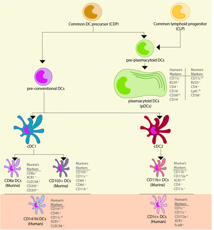
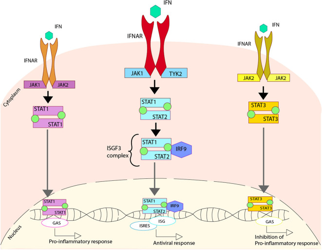
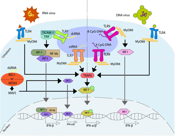
Dendritic cells (DCs) are a type of innate immune cells with major relevance in the establishment of an adaptive response, as they are responsible for the activation of lymphocytes. Since their discovery, several reports of their role during infectious diseases have been performed, highlighting their functions and their mechanisms of action. DCs can be categorized into different subsets, and each of these subsets expresses a wide arrange of receptors and molecules that aid them in the clearance of invading pathogens. Interferon (IFN) is a cytokine -a molecule of protein origin- strongly associated with antiviral immune responses. This cytokine is secreted by different cell types and is fundamental in the modulation of both innate and adaptive immune responses against viral infections. Particularly, DCs are one of the most important immune cells that produce IFN, with type I IFNs (α and β) highlighting as the most important, as they are associated with viral clearance. Type I IFN secretion can be induced via different pathways, activated by various components of the virus, such as surface proteins or genetic material. These molecules can trigger the activation of the IFN pathway trough surface receptors, including IFNAR, TLR4, or some intracellular receptors, such as TLR7, TLR9, and TLR3. Here, we discuss various types of dendritic cells found in humans and mice; their contribution to the activation of the antiviral response triggered by the secretion of IFN, through different routes of the induction for this important antiviral cytokine; and as to how DCs are involved in human infections that are considered highly frequent nowadays.

Keywords: IFN; antiviral response; dendritic cells; immune response; viruses.


Figures

Figure 1
Figure 1
Dendritic cell subsets. Dendritic cells (DCs) are derived from a common myeloid precursor, from which two precursors can be developed. The first precursor corresponds to the pre-conventional DCs and in murine models they can become conventional DCs (cDCs) type 1 and 2, and within these cells there are different subtypes of DCs. In the cDC1 found two subtypes of cells can be found: CD8+ DCs which has the markers CD8+, XCR1+, CLEC9A+, CD205+, and CD207+, and CD103+ DCs that has the markers CD103+, CD11c+, CD80+, CD86+, and CD11b+. The functional homolog of these cells in human are the CD141hi DCs, and their markers correspond to CD141hi, CD45+, CD11clo, XCR1+, and CLEC9A+. In the cDC2 subsets are comprised the CD11b+ DCs which has the markers CD11b+, CD172ahi, XCR1lo/−, CD4+, and CD11c+. The functional homolog of these cells in human are the CD1c+ DCs, and their markers are CD1c+, CD11c+, CD172a+, XCR1, and FcεRI+. The second precursor corresponds to the pre-plasmacytoid DCs that can become plasmacytoid DCs (pDCs), and in murine models its markers correspond to CD11lo, B220+, CD4+, Ly6Chi, and CD38+, while in human, their markers correspond to CD11, B220+, CD4+, CD11a+, and CD38lo.
Figure 2
Figure 2
Regulation of type I IFN due to the activation of IFN receptors. Upon activation of the IFNAR receptor induced by the cytokine IFN, three different pathways can be activated. One pathway involves the phosphorylation of the IFNAR cause by Janus Kinase 1 (JAK1) and Tyrosine Kinase 2 (TYK2), which generates the phosphorylation of both STAT1 and STAT2 that come together to form a heterodimer. This heterodimer interacts with IRF9, forming the ISGF3 complex that is translocated to the nucleus where it binds to DNA activating the regions of ISG and ISRES, promoting an antiviral response. An alternative pathway involves the phosphorylation of the IFNAR caused by JAK1 and JAK2, which produces the phosphorylation of STAT1 and two of them come together in order to form a homodimer. This homodimer is able to translocate to the nucleus where it binds to DNA, activating the regions of GAS, and promoting a pro-inflammatory response. Another alternative pathway involves the phosphorylation of the IFNAR, caused by JAK2, which generates the phosphorylation of STAT3, and two of these come together in order to form a homodimer. This homodimer is able to translocate to the nucleus where it binds to DNA, activating the regions of GAS and promoting the inhibition of the pro-inflammatory response.
Figure 3
Figure 3
Regulation of type I IFN due to the activation of Toll-like receptors. Upon activation of Toll-like receptor (TLR) induced directly by viruses, different pathways can be activated. TLR7 recognize ssRNA, promoting the binding with MYD88, that will later interact with TRAF6, activating IRF7 which is translocated to the nucleus where it binds to DNA, promoting the release of IFN-α/β. TLR3 recognize dsRNA, promoting the interaction with TICAM-1 and TRIF, that activates AP-1, NF-κB, and IRF3, which are translocated to the nucleus, where they bind to DNA, promoting the release of IFN-β. The dsRNA can activate an alternative pathway, which can be sensed by either retinoic acid-inducible gene I (RIG-I) or by melanoma differentiation-associated protein 5 (MDA5). These receptors activate mitochondrial antiviral-signaling protein (MAVS), and by doing so they promote the IRF3 and/or IRF7 activation, and later translocation into the nucleus where they bind to DNA, promoting the release of IFN-α/β. There are two ways of activating TLR9, one is through the recognition of a type A CpG-DNA, which activates the pathway mediated by MYD88, similar to TLR7, while the other one involves the recognition of a type B CpG-DNA, where TLR9 binds with MYD88, activating IRF1, which is then translocated into the nucleus where it binds to DNA promoting the release of IFN-β. On the other hand, TLR4 is able to recognize viral peptides, activating a MYD88 -depending pathway similar to TLR7.

References

    1. Banchereau J, Steinman RM. Dendritic cells and the control of immunity. Nature. (1998) 392:245–52. 10.1038/32588 - DOI - PubMed
    1. Pollara G, Kwan A, Newton PJ, Handley ME, Chain BM, Katz DR. Dendritic cells in viral pathogenesis: protective or defective? Int J Exp Pathol. (2005) 86:187–204. 10.1111/j.0959-9673.2005.00440.x - DOI - PMC - PubMed
    1. Banchereau J, Briere F, Caux C, Davoust J, Lebecque S, Liu YJ, et al. Immunobiology of dendritic cells. Annu Rev Immunol. (2000) 18:767–811. 10.1146/annurev.immunol.18.1.767 - DOI - PubMed
    1. Steinman RM, Cohn ZA. Identification of a novel cell type in peripheral lymphoid organs of mice. I. Morphology, quantitation, tissue distribution. J Exp Med. (1973) 137:1142–62. 10.1084/jem.137.5.1142 - DOI - PMC - PubMed
    1. Buchta CM, Bishop GA. Toll-like receptors and B cells: functions and mechanisms. Immunol Res. (2014) 59:12–22. 10.1007/s12026-014-8523-2 - DOI - PubMed

Publication types