. 2020 Jul 24;6(8):e589.
doi: 10.1097/TXD.0000000000001012. eCollection 2020 Aug.

Cellular Mechanisms of Rejection of Optic and Sciatic Nerve Transplants: An Observational Study

Affiliations

Cellular Mechanisms of Rejection of Optic and Sciatic Nerve Transplants: An Observational Study

Merve Yonar et al. Transplant Direct. .

Abstract

Background: Organ transplantation is a standard therapeutic strategy for irreversible organ damage, but the utility of nerve transplantation remains generally unexplored, despite its potential benefit to a large patient population. Here, we aimed to establish a feasible preclinical mouse model for understanding the cellular mechanisms behind the rejection of peripheral and optic nerves.

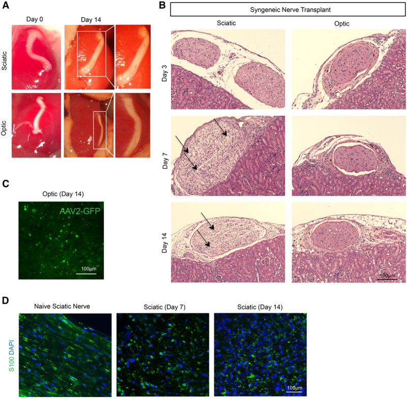
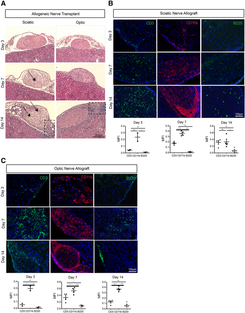
Methods: We performed syngenic and allogenic transplantation of optic and sciatic nerves in mice by inserting the nerve grafts inside the kidney capsule, and we assessed the allografts for signs of rejection through 14 d following transplantation. Then, we assessed the efficacy of CTLA4 Ig, Rapamycin, and anti-CD3 antibody in suppressing immune cell infiltration of the nerve allografts.

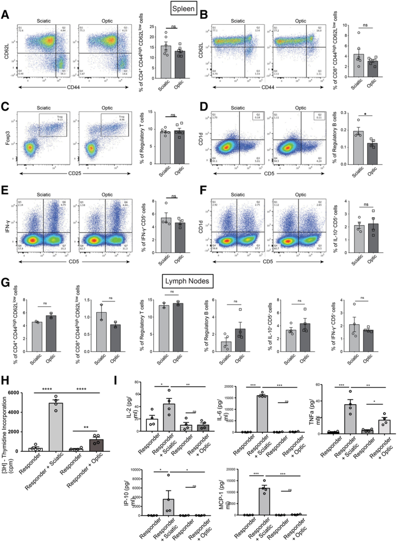
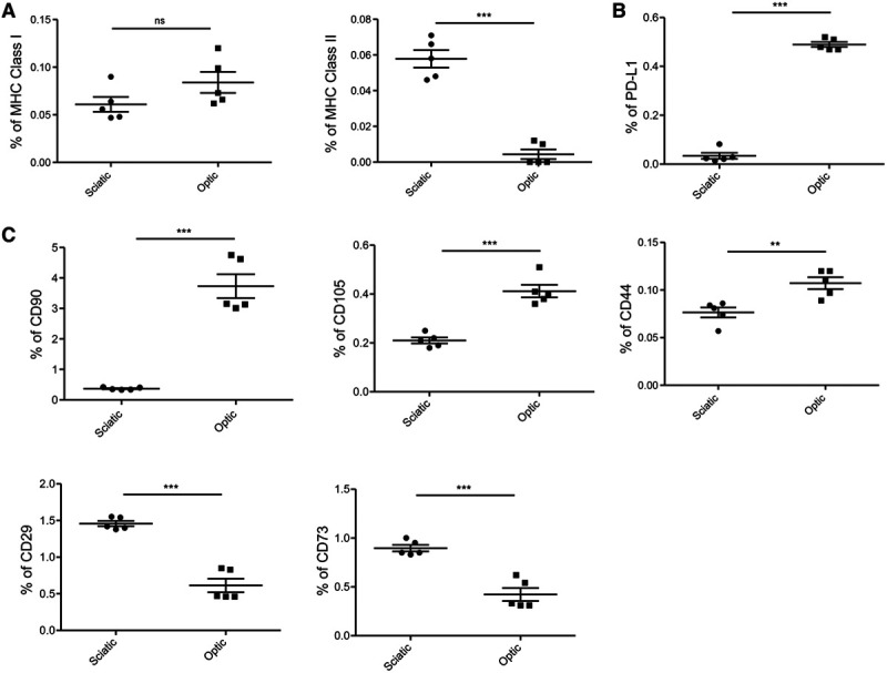
Results: By 3 d posttransplantation, both sciatic and optic nerves transplanted from BALB/c mice into C57BL/6J recipients contained immune cell infiltrates, which included more CD11b+ macrophages than CD3+ T cells or B220+ B cells. Ex vivo immunogenicity assays demonstrated that sciatic nerves demonstrated higher alloreactivity in comparison with optic nerves. Interestingly, optic nerves contained higher populations of anti-inflammatory PD-L1+ cells than sciatic nerves. Treatment with anti-CD3 antibody reduced immune cell infiltrates in the optic nerve allograft, but exerted no significant effect in the sciatic nerve allograft.

Conclusions: These findings establish the feasibility of a preclinical allogenic nerve transplantation model and provide the basis for future testing of directed, high-intensity immunosuppression in these mice.


Conflict of interest statement

The authors declare no conflicts of interest.

Figures

FIGURE 1.
FIGURE 1.
Autologous optic and sciatic nerve transplantation. A, Representative photographs demonstrate a macroscopic appearance of nerve grafts on d 0 and 14 following autologous transplantation of sciatic and optic nerves between genetically identical C57BL/6J mice. B, Light micrographs of H&E-stained sciatic and optic nerve autografts on d 3, 7, and 14. Arrows indicate allograft regions affected by vacuolization. Images are representative of 5 independent experiments (n = 5). C, Following intraophthalmic injection of GFP-labeled adeno-associated virus-2 before procurement of optic nerve, fluorescence micrograph of optic nerve autograft on d 14 demonstrates the presence of GFP, confirming the presence of axons. D, Fluorescence micrograph of S100+ Schwann cells (green) in naïve sciatic nerve and sciatic autografts on d 7 and 14 demonstrate persistence of S100+ signal. AAV2-GFP, GFP-labeled adeno-associated virus 2; H&E, hematoxylin and eosin.
FIGURE 2.
FIGURE 2.
Allogeneic optic and sciatic nerve transplantation and characterization of immune cells. A, H&E images of sciatic and optic grafts on d 3, 7, and 14 after allogeneic transplantation between BALB/c and C57BL/6J mouse. Arrows indicate allograft regions affected by vacuolization, and boxes indicate areas of immune cell infiltration. Images are representative of 5 independent experiment (n = 5). B, Representative fluorescence micrographs of CD3+ T cells (green), B220+ B cells (green), and CD11b+ cells (red) in sciatic nerve allografts at d 3, 7, and 14 following transplantation from BALB/c mouse into C57BL6/J recipient (n = 2 at d 3, n = 4 at d 7 and 14). Comparisons of MFI of CD3+, B220+, and CD11b+ signals in the sciatic nerve allograft at d 3 (n = 2), d 7 (n = 4), and d 14 (n = 4). C, Representative fluorescence micrographs of CD3+ T cells (green), B220+ B cells (green), and CD11b+ macrophages (red) in optic nerve allografts at d 3, 7, and 14 following transplantation from BALB/c mouse into C57BL6/J recipient (n = 2 at d 3, n = 4 at d 7 and 14). Comparisons of MFI of CD3+, B220+, and CD11b+ signals in the optic nerve allograft at d 3 (n = 2), d 7 (n = 4), and d 14 (n = 4). Data are shown as mean ± SEM; student t-test. H&E, hematoxylin and eosin; MFI, mean fluorescence intensity.
FIGURE 3.
FIGURE 3.
Assessment of alloimmune response in secondary lymphoid organs of nerve allograft recipient mice. A–F, Comparisons of the (A) CD44high CD62Llow CD4+ effector memory T cell (15.77% vs 13.22%; P = 0.2454), (B) CD44high CD62Llow CD8+ effector memory T cell (4.405% vs 3.092%; P = 0.2155), (C) CD25+ FOXP3+ regulatory T cell (9.655% vs 9.332%; P = 0.6666), (D) B220+ CD1d+ CD5+ regulatory B cell (0.1950% vs 0.1250%; P = 0.0304), (E) IFN-γ+ CD5+ cell (5.423% vs 4.658%; P = 0.3924), and (F) IL-10+ CD5+ cell (2.128% vs 2.250%; P = 0.8137) populations between the spleens of mice that have received either optic or sciatic nerve allografts. G, Comparisons of the CD44high CD62Llow CD4+ effector memory T cell (4.580% vs 5.570%; P = 0.1209), CD44high CD62Llow CD8+ effector memory T cell (1.140% vs 0.7850%; P = 0.2662), CD25+ FOXP3+ regulatory T cell (13.40% vs 14.00%; P = 0.5577), B220+ CD1d+ CD5+ regulatory B cell (1.160% vs 2.648%; P = 0.1392), IFN-γ+ CD5+ cell (2.113% vs 1.685%; P = 0.4818), and IL-10+ CD5+ cell (3.355% vs 4.350%; P = 0.2963) populations between the lymph nodes of mice that have received either optic or sciatic nerve allografts. Data were collected from 4 independent experiments and shown as mean ± SEM; student t-test. H, Mixed lymphocyte reaction (MLR) assay with splenocytes from transplanted mice (sciatic or optic) and nerve cells (sciatic and optic) from donor mice (BALB/c). The splenocytes from the sciatic nerve-transplanted group showed significantly higher proliferation compared with the optic nerve-transplanted group (responder+sciatic vs responder+optic, 4967 ± 312.5 vs 1237 ± 222.5, ***P < 0.001, n = 4/group). I, Luminex assay showed significantly higher inflammatory cytokines and chemokines in the media taken from the sciatic nerve-transplanted group in comparison with the optic nerve-transplanted group (responder+sciatic vs responder+optic, 44.6 ± 8.6 vs 10.4 ± 3.1, **P < 0.01 for IL-2, 16 065 ± 463.1 vs 213.1 ± 56.9, ***P < 0.001 for IL-6, 35.8 ± 5.8 vs 16.9 ± 2.9, **P < 0.01 for TNFα, 3605 ± 1828 vs 45.5 ± 13.8, *P < 0.05 for IP-10, 11 793 ± 1196 vs 218.9 ± 64.2, ***P < 0.001 for MCP-1, n = 4/group). MCP-1, monocyte chemoattractant protein-1.
FIGURE 4.
FIGURE 4.
Assessment of sciatic and optic nerve by flow cytometry. A, Comparison of percentages of MHC Class I and MHC Class II (sciatic vs optic, 0.06% vs 0.08%, P = ns for MHC Class I, 0.06% vs 0.004%, ***P < 0.001 for MHC Class II, n = 5/group). B, Optic nerve showed significantly higher PD-L1 population (sciatic vs optic, 0.03% vs 0.4%, ***P < 0.001, n = 5/group). C, Optic nerve showed significantly higher population of CD90, CD105, and CD44 compared with sciatic nerve (sciatic vs optic, 0.3% vs 3.7%, ***P < 0.001 for CD90, 0.2% vs 0.4%, ***P < 0.001 for CD105, 0.07% vs 0.11%, **P < 0.01 for CD44, n = 5/group). CD73 and CD29 were less in optic nerve compared with sciatic nerve (sciatic vs optic, 0.4% vs 0.2%, ***P < 0.001 for CD73, 0.52% vs 0.24%, ***P < 0.001 for CD29, n = 5/group). MHC, major histocompatibility complex.
FIGURE 5.
FIGURE 5.
Identification of effective immunosuppressive regimens for prevention of nerve allograft rejection. A, Representative light micrographs of H&E-stained nerve allografts demonstrate the comparison between the histologic appearance at d 14 of the untreated control group and nerve allografts from mice treated with CTLA4 Ig or Rapamycin. Arrows indicate allograft regions affected by vacuolization, and boxes indicate areas of immune cell infiltration. B, Comparisons of cellular infiltration scores at d 14 between sciatic nerve allografts or optic nerve allografts from untreated mice and those from mice treated with CTLA4 Ig or Rapamycin. C, Representative light micrographs of H&E-stained heart allografts demonstrate a comparison between the histologic appearance of the untreated control group and allografts from mice treated with CTLA4 Ig. D, Representative fluorescence micrographs of CD3+ T cell (green), B220+ B cell (green), and CD11b+ macrophage (red) populations in optic nerve allograft at d 7 following treatment with anti-CD3 and sciatic nerve allograft at d 14 following treatment with anti-CD3. E, Comparison of MFI of CD3+ (0.3883% vs 0.0084%, **P < 0.01; n = 3), B220+ (0.0970% vs 0.0142%, *P < 0.05; n = 3), and CD11b+ signals (0.5763% vs 0.3527%, *P < 0.05; n = 3) between sciatic nerve allografts from untreated mice and those from mice treated with anti-CD3. F, Comparison of MFI of CD3+ (0.2874% vs 0.1457%, P = ns; n = 3), B220+ (0.0342% vs 0.0372%, P = ns; n = 3), and CD11b+ signals (0.2488% vs 0.1537%, P = ns; n = 3) between optic nerve allografts from untreated mice and those from mice treated with anti-CD3. Data are shown as mean ± SEM, student t-test. H&E, hematoxylin and eosin; MFI, mean fluorescence intensity.

References

    1. Park J, Lin H-Y, Assaker JP, et al. Integrated kidney exosome analysis for the detection of kidney transplant rejection. ACS Nano. 2017; 11:11041–11046. doi:10.1021/acsnano.7b05083 - PMC - PubMed
    1. Taylor DO, Edwards LB, Mohacsi PJ, et al. The registry of the International Society for Heart and Lung Transplantation: twentieth official adult heart transplant report–2003. J Heart Lung Transplant. 2003; 22:616–624. doi:10.1016/s1053-2498(03)00186-4 - PubMed
    1. Pomahac B, Bueno EM, Sisk GC, et al. Current principles of facial allotransplantation: the Brigham and Women’s Hospital experience. Plast Reconstr Surg. 2013; 131:1069–1076. doi:10.1097/PRS.0b013e3182865cd3 - PMC - PubMed
    1. Elliott RM, Tintle SM, Levin LS. Upper extremity transplantation: current concepts and challenges in an emerging field. Curr Rev Musculoskelet Med. 2014; 7:83–88. doi:10.1007/s12178-013-9191-x - PMC - PubMed
    1. Benowitz LI, Yin Y. Optic nerve regeneration. Arch Ophthalmol. 2010; 128:1059–1064. doi:10.1001/archophthalmol.2010.152 - PMC - PubMed