. 2020 Jun 25:24:100434.
doi: 10.1016/j.eclinm.2020.100434. eCollection 2020 Jul.

Megakaryocytes and platelet-fibrin thrombi characterize multi-organ thrombosis at autopsy in COVID-19: A case series

Affiliations

Megakaryocytes and platelet-fibrin thrombi characterize multi-organ thrombosis at autopsy in COVID-19: A case series

Amy V Rapkiewicz et al. EClinicalMedicine. .

Abstract

Background: There is increasing recognition of a prothrombotic state in COVID-19. Post-mortem examination can provide important mechanistic insights.

Methods: We present a COVID-19 autopsy series including findings in lungs, heart, kidneys, liver, and bone, from a New York academic medical center.

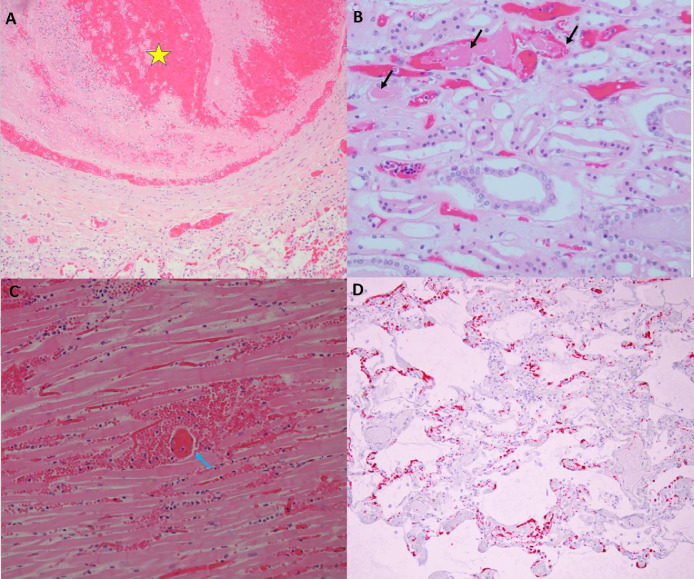
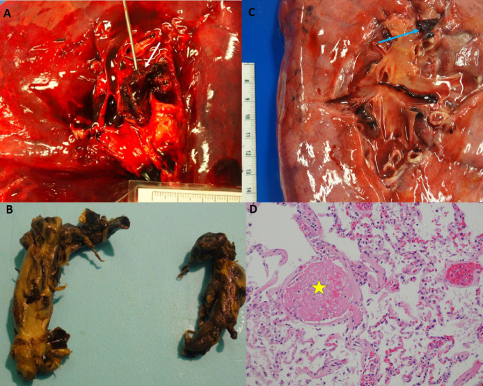
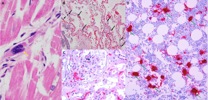
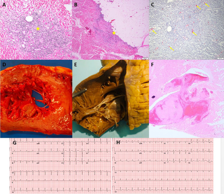
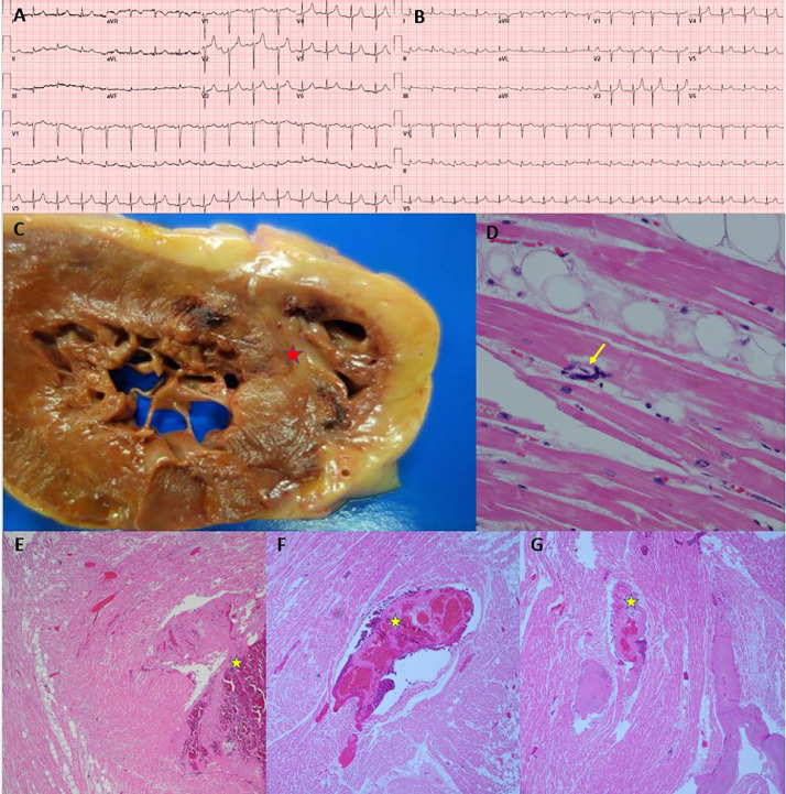
Findings: In seven patients (four female), regardless of anticoagulation status, all autopsies demonstrated platelet-rich thrombi in the pulmonary, hepatic, renal, and cardiac microvasculature. Megakaryocytes were seen in higher than usual numbers in the lungs and heart. Two cases had thrombi in the large pulmonary arteries, where casts conformed to the anatomic location. Thrombi in the IVC were not found, but the deep leg veins were not dissected. Two cases had cardiac venous thrombosis with one case exhibiting septal myocardial infarction associated with intramyocardial venous thrombosis, without atherosclerosis. One case had focal acute lymphocyte-predominant inflammation in the myocardium with no virions found in cardiomyocytes. Otherwise, cardiac histopathological changes were limited to minimal epicardial inflammation (n = 1), early ischemic injury (n = 3), and mural fibrin thrombi (n = 2). Platelet-rich peri‑tubular fibrin microthrombi were a prominent renal feature. Acute tubular necrosis, and red blood cell and granular casts were seen in multiple cases. Significant glomerular pathology was notably absent. Numerous platelet-fibrin microthrombi were identified in hepatic sinusoids. All lungs exhibited diffuse alveolar damage (DAD) with a spectrum of exudative and proliferative phases including hyaline membranes, and pneumocyte hyperplasia, with viral inclusions in epithelial cells and macrophages. Three cases had superimposed acute bronchopneumonia, focally necrotizing.

Interpretation: In this series of seven COVID-19 autopsies, thrombosis was a prominent feature in multiple organs, in some cases despite full anticoagulation and regardless of timing of the disease course, suggesting that thrombosis plays a role very early in the disease process. The finding of megakaryocytes and platelet-rich thrombi in the lungs, heart and kidneys suggests a role in thrombosis.

Funding: None.

Keywords: “Autopsy”; “COVID-19″; “Megakaryocyte”; “Thrombosis”.


Conflict of interest statement

Dr. Rapkiewicz, Dr. Mai, Dr. Pittaluga, Dr. Thomas, Dr. Kleiner, Dr. Adler and Dr. Gasmi have nothing to disclose. Dr. Carsons reports grants from Novartis and grants from GSK, outside the submitted work. Dr. Berger reports grants from National Heart, Lung and Blood Institute, during the conduct of the study; grants from Astrazeneca, other from Jannsen, other from Amgen, outside the submitted work. Dr. Charytan reports personal fees from PLC Medical, grants from bioporto, personal fees from Merck, grants and personal fees from NovoNordisk, grants and personal fees from Jannssen, grants and personal fees from Gilead, personal fees from AstraZeneca, grants and personal fees from Medtronic, grants and personal fees from Amgen, personal fees from Fresenius, personal fees from GSK, outside the submitted work. Dr. Hochman was supported in part by the NYU CTSA grant UL1TR001445, from the National Center for Advancing Translational Sciences (NCATS) during the conduct of the study; grants from NHLBI, other from AstraZeneca Pharmaceuticals LLC, other from Arbor Pharmaceuticals LLC, non-financial support from Abbott Vascular, non-financial support from Medtronic Inc, non-financial support from St. Jude Medical Inc, non-financial support from Volcano Corp, non-financial support from Merck Sharp & Dohme Corp, non-financial support from Omron Healthcare Inc, non-financial support from Amgen Inc, outside the submitted work. Dr. Reynolds reports non-financial support from Abbott Vascular, non-financial support from Siemens, non-financial support from Biotelemetry Inc outside the submitted work.

Figures

Fig. 1
Fig. 1
Thrombosis in multiple organs at autopsy. Panel A, Case 3, 20x magnification of hematoxylin and eosin stained section, medium vessel pulmonary artery thrombus (yellow star) with lines of Zahn and fibrin thrombus without organization in a smaller arteriole (yellow arrow). The adjacent lung shows similar temporal relationship with exudative phase of diffuse alveolar damage with hyaline membrane formation. Panel B, Case 2, 20x magnification of hematoxylin and eosin stained section of medulla of the kidney with fibrin microthrombi in the peritubular capillaries (black arrows). The tubular epithelium shows detachment from the basement membrane with vacuolization as well as granular debris within the tubular lumen. Panel C, Case 7, 20x magnification of hematoxylin and eosin stained section. Cardiac tissue with fibrin thrombus in a perforating vein (blue arrow) associated with a myocardial infarction showing myocardial necrosis, which was transmural, and neutrophilic infiltrates. Panel D, Case 2, 10x magnification of hematoxylin and eosin stained section. Platelet microthrombi highlighted by CD61 immunohistochemical stain within the microvasculature of the lung. (For interpretation of the references to color in this figure legend, the reader is referred to the web version of this article.)
Fig. 2
Fig. 2
Pulmonary thrombi. Panel A: Case 4, Large occlusive pulmonary thrombus within the right main pulmonary artery (white arrow). Panel B: Case 4, Thrombus easily removed showing a predominately white thrombus that is molded in the anatomic pattern of the pulmonary vasculature. Panel C: Case 5, Loosely adherent pulmonary thrombus within a segmental pulmonary artery of the left lung (blue arrow). Panel D: Case 5, H&E 40x, microscopic image of the lung showing changes of diffuse alveolar damage as well as a fibrin microthrombus (yellow star). (For interpretation of the references to color in this figure legend, the reader is referred to the web version of this article.)
Fig. 3
Fig. 3
Megakaryocytes in the heart, kidney, lung and bone marrow. Panel A: Case 4, 40x H&E stain, Heart with early ischemic changes and megakaryocyte (yellow arrow) and fibrin thrombus within the lumen of a venule. Panel B: Case 2, 20x H&E stain, Lung with pulmonary edema and early bronchopneumonia with multiple megakaryocytes (black arrows) within the alveolar capillaries. Panel C: Case 4, 20x H&E stain, Megakaryocyte within the capillary loop of a glomerulus (blue arrow). Panel D: Case 3, Hypercellular marrow for age with trilineage hematopoiesis. There is an increased number of megakaryocytes with focal clustering (green arrows). Myeloid elements show only focal evidence of progressive maturation (left shift). Megakaryocytes and platelets are highlighted by CD61 staining. (For interpretation of the references to color in this figure legend, the reader is referred to the web version of this article.)
Fig. 4
Fig. 4
Myocardial Inflammation and Right Ventricular Mural Thrombus (Case 4). Panels A and B: H&E 10x, Myocardium with a focal chronic inflammatory infiltrate composed of lymphocytes with associated myocardial necrosis in the epicardial region (yellow stars). Panel C: Platelet microthrombi highlighted by CD61 immunohistochemical stain within the microvasculature of the heart (yellow arrows), 10x. Panel D: Gross image of a cut section of the heart showing transmural diffuse pallor of the left ventricle. Panel E: Right ventricle of the heart with mural white thrombus within the trabeculae carnae. Panel F: Microscopic findings in the right ventricle of the heart with mural white thrombus within the trabeculae carnae. Panel G: Admission ECG shows normal sinus rhythm without significant ST segment abnormality.
Fig. 5
Fig. 5
Myocardial Infarction due to Venous Thrombosis (Case 7). Panel A: Admission ECG showing normal sinus rhythm. Panel B: ECG after intubation (day of death), showing sinus rhythm with premature atrial contraction and inferolateral ST segment elevation. Panel C: Cut section of left and right ventricle showing pallor with peripheral hemorrhage rim at the juncture of the posterior ventricles and interventricular septum (red star). Panel D: Microscopic section showing megakaryocyte within a small vessel in the myocardium (yellow arrow). Panels E, F, G: H&E sections of the right and left ventricle with intramyocardial venous thrombosis (yellow stars), fibrin microthrombi and varying degrees of infarction.

References

    1. Wang D., Hu B., Hu C. Clinical characteristics of 138 hospitalized patients with 2019 novel coronavirus-infected pneumonia in Wuhan, China. JAMA. 2020;323(11):1061–1069. - PMC - PubMed
    1. Chen T., Wu D., Chen H. Clinical characteristics of 113 deceased patients with coronavirus disease 2019: retrospective study. BMJ. 2020;368:m1091. - PMC - PubMed
    1. Guo T., Fan Y., Chen M. Cardiovascular Implications of Fatal Outcomes of Patients With Coronavirus Disease 2019 (COVID-19) JAMA Cardiol. 2020:e201017. - PMC - PubMed
    1. Shi S., Qin M., Shen B. Association of Cardiac Injury With Mortality in Hospitalized Patients With COVID-19 in Wuhan, China. JAMA Cardiol. 2020 Published online March 25, 2020. - PMC - PubMed
    1. Bangalore S., Sharma A., Slotwiner A. ST-Segment Elevation in Patients with Covid-19 - A Case Series. N Engl J Med. 2020 NEJMc2009020. - PMC - PubMed