

White Matter Microstructural Abnormalities in the Broca's-Wernicke's-Putamen "Hoffman Hallucination Circuit" and Auditory Transcallosal Fibers in First-Episode Psychosis With Auditory Hallucinations
- PMID: 32766733
- PMCID: PMC7825092
- DOI: 10.1093/schbul/sbaa105
White Matter Microstructural Abnormalities in the Broca's-Wernicke's-Putamen "Hoffman Hallucination Circuit" and Auditory Transcallosal Fibers in First-Episode Psychosis With Auditory Hallucinations
Abstract
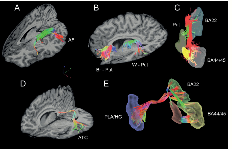
Background: Functional connectivity abnormalities between Broca's and Wernicke's areas and the putamen revealed by functional magnetic resonance imaging (fMRI) are related to auditory hallucinations (AH). In long-term schizophrenia, reduced white matter structural integrity revealed by diffusion imaging in left arcuate fasciculus (connecting Broca's and Wernicke's areas) is likely related to AH. The structural integrity of connections with putamen and their relation to AH are unknown. Little is known about this relationship in first-episode psychosis (FEP), although auditory transcallosal connections were reported to play a role. White matter in the Broca's-Wernicke's-putamen language-related circuit and auditory transcallosal fibers was examined to investigate associations with AH in FEP.
Methods: White matter connectivity was measured in 40 FEP and 32 matched HC using generalized fractional anisotropy (gFA) derived from diffusion spectrum imaging (DSI).
Results: FEP and HC did not differ in gFA in any fiber bundle. In FEP, AH severity was significantly inversely related to gFA in auditory transcallosal fibers and left arcuate fasciculus. Although the right hemisphere arcuate fasciculus-AH association did not attain significance, the left and right arcuate fasciculus associations were not significantly different.
Conclusions: Despite overall normal gFA in FEP, AH severity was significantly related to gFA in transcallosal auditory fibers and the left hemisphere connection between Broca's and Wernicke's areas. Other bilateral tracts' gFA were weakly associated with AH. At the first psychotic episode, AH are more robustly associated with left hemisphere arcuate fasciculus and interhemispheric auditory fibers microstructural deficits, likely reflecting mistiming of information flow between language-related cortical centers.
Keywords: Broca’s area; Wernicke’s area; arcuate fasciculus; auditory hallucination; first-episode psychosis; putamen; transcallosal fibers.
© The Author(s) 2020. Published by Oxford University Press on behalf of the Maryland Psychiatric Research Center. All rights reserved. For permissions, please email: journals.permissions@oup.com.
Figures

References
-
- Binder JR, Frost JA, Hammeke TA, Rao SM, Cox RW. Function of the left planum temporale in auditory and linguistic processing. Brain. 1996;119 (Pt 4):1239–1247. - PubMed
-
- Smith EE, Jonides J, Koeppe RA. Dissociating verbal and spatial working memory using PET. Cereb Cortex. 1996;6(1):11–20. - PubMed
-
- Poldrack RA, Wagner AD, Prull MW, Desmond JE, Glover GH, Gabrieli JD. Functional specialization for semantic and phonological processing in the left inferior prefrontal cortex. Neuroimage. 1999;10(1):15–35. - PubMed
Publication types
MeSH terms
Grants and funding
LinkOut - more resources
Full Text Sources
Medical
