. 2020 Oct 9;295(41):14015-14024.
doi: 10.1074/jbc.RA120.013440. Epub 2020 Aug 7.

The inhibition of cellular toxicity of amyloid-β by dissociated transthyretin

Affiliations

The inhibition of cellular toxicity of amyloid-β by dissociated transthyretin

Qin Cao et al. J Biol Chem. .

Abstract

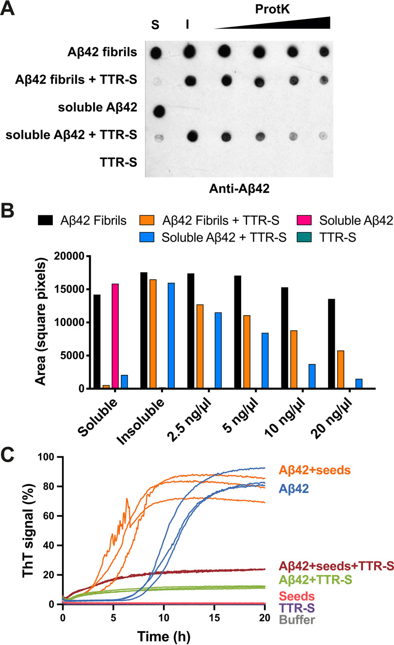
The protective effect of transthyretin (TTR) on cellular toxicity of β-amyloid (Aβ) has been previously reported. TTR is a tetrameric carrier of thyroxine in blood and cerebrospinal fluid, the pathogenic aggregation of which causes systemic amyloidosis. However, studies have documented a protective effect of TTR against cellular toxicity of pathogenic Aβ, a protein associated with Alzheimer's disease. TTR binds Aβ, alters its aggregation, and inhibits its toxicity both in vitro and in vivo In this study, we investigate whether the amyloidogenic ability of TTR and its antiamyloid inhibitory effect are associated. Using protein aggregation and cytotoxicity assays, we found that the dissociation of the TTR tetramer, required for its amyloid pathogenesis, is also necessary to prevent cellular toxicity from Aβ oligomers. These findings suggest that the Aβ-binding site of TTR may be hidden in its tetrameric form. Aided by computational docking and peptide screening, we identified a TTR segment that is capable of altering Aβ aggregation and toxicity, mimicking TTR cellular protection. EM, immune detection analysis, and assessment of aggregation and cytotoxicity revealed that the TTR segment inhibits Aβ oligomer formation and also promotes the formation of nontoxic, nonamyloid amorphous aggregates, which are more sensitive to protease digestion. Finally, this segment also inhibits seeding of Aβ catalyzed by Aβ fibrils extracted from the brain of an Alzheimer's patient. Together, these findings suggest that mimicking the inhibitory effect of TTR with peptide-based therapeutics represents an additional avenue to explore for the treatment of Alzheimer's disease.

Keywords: Alzheimer's disease; amyloid; amyloid-beta (AB); cytotoxicity; neuroprotection; neuroscience; peptide; transthyretin.


Conflict of interest statement

Conflict of interest—L. S. is a consultant for ADRx, Inc.

Figures

Figure 1.
Figure 1.
The inhibition of Aβ42 cytotoxicity by TTR depends on the aggregation propensity of each TTR variant. A, aggregation assay of three TTR variants followed by immunodot blot of insoluble fractions collected prior to incubation or after 1, 2, and 4 days of incubation at 37° C, as labeled. All samples were treated equally and onto the same membrane. Cropping was applied for ethetical reasons. B, electron micrographs of aggregated TTR variants after 4 days of incubation. Scale bar, 200 nm. C, size-exclusion chromatography of soluble TTR variants at pH 7.4. D, cytotoxicity assay of Aβ42 in the presence of TTR variants at different stages of aggregation, followed by MTT reduction. A 5-fold molar excess of soluble TTR (50 μm, considering monomeric concentration) and aggregated samples collected after 1, 2 and 4 days of incubation were added to soluble 10 μm Aβ42 and incubated overnight. The samples were added to HeLa cells, and MTT reduction was measured after 24 h. Buffer-treated cells were considered 100% viability and used for normalization (n = 3). All replicates are shown. Error bars, S.D. **, p ≤ 0.005; ***, p ≤ 0.0005. N.D., significance not detected. These results suggest that the dissociation of the tetrameric TTR structure precedes Aβ42 cytotoxicity protection, whereas aggregated TTR does not exert any effect.
Figure 2.
Figure 2.
Protein–protein docking of TTR monomer with three KLVFFA polymorphs. Protein–protein docking was performed using monomeric TTR and three KLVFFA polymorphs identified previously (17): PDB codes 2y2a, 2y21, and 3ow9. A, monomeric TTR obtained from chain A of the model of PDB code 4TLT (18) is shown in gray as a secondary structure. On the left is shown a view down the hydrophobic pocket. On the right is a lateral view. B, fibrillar structures of KLVFFA from PDB codes 2y2a (left panel), 2y21 (middle panel), and 3ow9 (right panel). Only one sheet of each KLVFFA polymer was included in the docking, shown in blue. The resulting docking models, shown in C, suggest that TTR may interact with Aβ42 through residues 105–117. Monomeric TTR is shown in gray with the segment TTR(105–117) in red. Top row, lateral view of interface. Bottom row, view down the binding interface. Residues involved in the interaction between monomeric TTR and KLVFFA are shown as sticks. Spheres represent the van der Waals radii of the side chain atoms of the tightly packed binding interface.
Figure 3.
Figure 3.
Effect of TTR-S on both soluble and fibrillar Aβ42. A, ThT fluorescence was measured from samples containing 10 μm Aβ42 in the presence and absence of a 3-fold molar excess of TTR-S. TTR-S was added before incubation (green) or after 18 h of incubation (red). TTR-S alone (purple) and buffer (not shown) were used as negative controls (n = 4). All replicates are shown. For a complete assessment of Aβ42 inhibition by TTR-derived peptides, see Fig. S4. B, electron micrographs of Aβ42 fibrils formed in the absence of inhibitor (top panel), Aβ42 amorphous aggregates formed when TTR-S was added after 18 h of incubation (middle panel), and Aβ42 amorphous aggregates formed when TTR-S was added prior to incubation (bottom). Scale bar, 400 nm. Insets, pictures of test tubes containing the same samples shown by EM but 10 times more concentrated. These pictures were taken after 30 min of incubation and reveal obvious precipitation upon addition of TTR-S. C, Congo red staining of samples shown in A under bright field (left panels) and polarized light (right panels). D, CD traces of Aβ42 fibrils, Aβ42 fibrils after addition of TTR-S, and soluble Aβ42 after addition of TTR-S. The table below shows the calculated percentage of various structural conformations: helix, β-stranded, and others (turns and unstructured). These analyses indicate that the TTR-derived peptide TTR-S inhibits Aβ42 aggregation and promotes the formation of nonamyloid amorphous aggregates.
Figure 4.
Figure 4.
TTR-S inhibition of Aβ42 cytotoxicity. A–C, cell viability assay followed by MTT reduction in which HeLa (A), PC12 (B), and SH-5YSY (C) cells were treated with 10 μm Aβ42 in the absence and the presence of increasing concentrations of peptides. Molar ratios of Aβ42 and peptides are labeled. As a negative control, cytotoxicity of peptides alone was also measured. Buffer-treated cells were considered 100% viability and used for normalization. Cell toxicity measured in the absence of peptides is marked with a red continuous line (mean) and dotted lines (S.D.; n = 3). All replicates are shown. Error bars, S.D. *, p ≤ 0.05; **, p ≤ 0.005; ***, p ≤ 0.0005. N.D., significance not detected. D, nondenaturing electrophoresis followed by immunoblot of Aβ42 before and after 16 h of incubation at 37 °C without and with TTR-S. Two gels were run to analyze Aβ42 before and after incubation and are shown in separate insets. The same samples were analyzed by dot blot with the oligomer specific A11 antibody (bottom panel). These results suggest that the inhibition of Aβ42 cytotoxicity by TTR-S results from the elimination of toxic oligomeric species.
Figure 5.
Figure 5.
Evaluation of the inhibitory effect of TTR-S by protease digestion and amyloid seeding. A, anti-Aβ42 immunodot blot of Aβ42 aggregates after proteinase K digestion. Soluble Aβ42 and preformed fibrils were incubated with TTR-S. Half of the sample was subjected to centrifugation, and soluble (S) and insoluble (I) fractions were collected. Increasing concentrations of proteinase K were added to the other half of the sample and incubated at 37° C for 1 h. The samples were analyzed by dot blot using E610 specific anti-Aβ42 antibody. As negative control, we included a sample with TTR-S alone. B, the signal was quantified using ImageJ and plotted as shown. This assay shows that Aβ42 amorphous aggregates generated after the addition of TTR-S are degraded by proteinase K more readily than Aβ42 fibrils. C, amyloid seeding assay followed by ThT signal. Soluble Aβ42 was incubated with AD ex vivo seeds in the presence or absence of TTR-S. Samples with only TTR-S, with only AD seeds, or with buffer were included as negative control. Note that the addition of TTR-S results in inhibition of Aβ42 seeding (n = 3). All replicates are shown.

References

    1. Schwarzman A. L., Gregori L., Vitek M. P., Lyubski S., Strittmatter W. J., Enghilde J. J., Bhasin R., Silverman J., Weisgraber K. H., Coyle P. K. (1994) Transthyretin sequesters amyloid β protein and prevents amyloid formation. Proc. Natl. Acad. Sci. U.S.A. 91, 8368–8372 10.1073/pnas.91.18.8368 - DOI - PMC - PubMed
    1. Roher A. E., Lowenson J. D., Clarke S., Woods A. S., Cotter R. J., Gowing E., and Ball M. J. (1993) β-Amyloid-(1–42) is a major component of cerebrovascular amyloid deposits: implications for the pathology of Alzheimer disease. Proc. Natl. Acad. Sci. U.S.A. 90, 10836–10840 10.1073/pnas.90.22.10836 - DOI - PMC - PubMed
    1. Burdick D., Soreghan B., Kwon M., Kosmoski J., Knauer M., Henschen A., Yates J., Cotman C., and Glabe C. (1992) Assembly and aggregation properties of synthetic Alzheimer's A4/β amyloid peptide analogs. J. Biol. Chem. 267, 546–554 - PubMed
    1. Stein T. D., Anders N. J., DeCarli C., Chan S. L., Mattson M. P., and Johnson J. A. (2004) Neutralization of transthyretin reverses the neuroprotective effects of secreted amyloid precursor protein (APP) in APPSW mice resulting in tau phosphorylation and loss of hippocampal neurons: support for the amyloid hypothesis. J. Neurosci. 24, 7707–7717 10.1523/JNEUROSCI.2211-04.2004 - DOI - PMC - PubMed
    1. Choi S. H., Leight S. N., Lee V. M., Li T., Wong P. C., Johnson J. A., Saraiva M. J., and Sisodia S. S. (2007) Accelerated Aβ deposition in APPswe/PS1deltaE9 mice with hemizygous deletions of TTR (transthyretin). J. Neurosci. 27, 7006–7010 10.1523/JNEUROSCI.1919-07.2007 - DOI - PMC - PubMed

Publication types

MeSH terms