. 2020 Aug 7;10(1):13314.
doi: 10.1038/s41598-020-69967-z.

Whole genome analysis reveals aneuploidies in early pregnancy loss in the horse

Affiliations

Whole genome analysis reveals aneuploidies in early pregnancy loss in the horse

Charlotte A Shilton et al. Sci Rep. .

Abstract

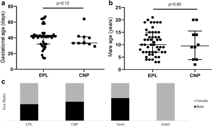
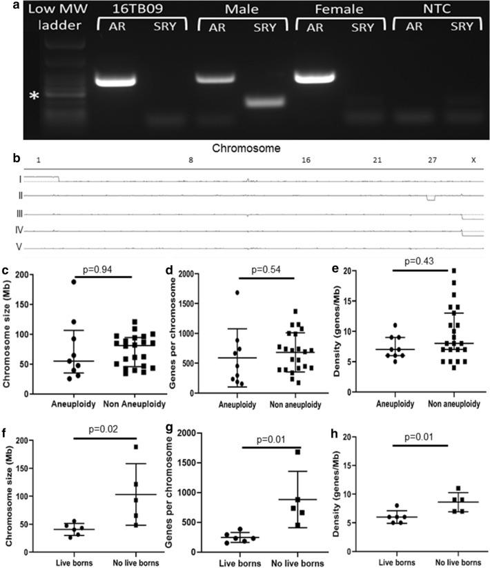
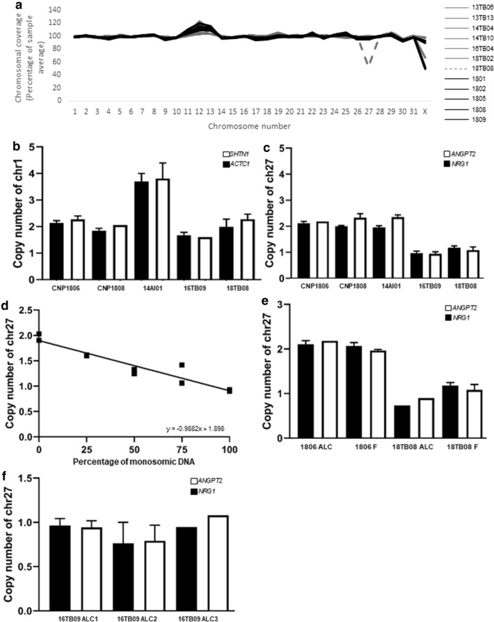
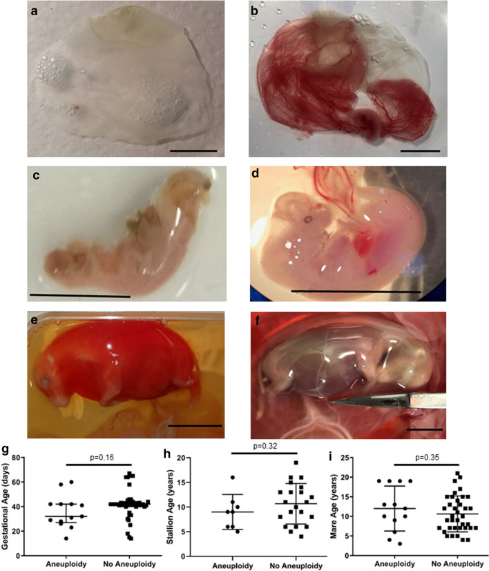
The first 8 weeks of pregnancy is a critical time, with the majority of pregnancy losses occurring during this period. Abnormal chromosome number (aneuploidy) is a common finding in human miscarriage, yet is rarely reported in domestic animals. Equine early pregnancy loss (EPL) has no diagnosis in over 80% of cases. The aim of this study was to characterise aneuploidies associated with equine EPL. Genomic DNA from clinical cases of spontaneous miscarriage (EPLs; 14-65 days of gestation) and healthy control placentae (various gestational ages) were assessed using a high density genotyping array. Aneuploidy was detected in 12/55 EPLs (21.8%), and 0/15 healthy control placentae. Whole genome sequencing (30X) and digital droplet PCR (ddPCR) validated results. The majority of these aneuploidies have never been reported in live born equines, supporting their embryonic/fetal lethality. Aneuploidies were detected in both placental and fetal compartments. Rodents are currently used to study how maternal ageing impacts aneuploidy risk, however the differences in reproductive biology is a limitation of this model. We present the first evidence of aneuploidy in naturally occurring equine EPLs at a similar rate to human miscarriage. We therefore suggest the horse as an alternative to rodent models to study mechanisms resulting in aneuploid pregnancies.


Conflict of interest statement

The authors declare no competing interests.

Figures

Figure 1
Figure 1
Description of sample population on the SNP array. No significant difference was found in the average (a) gestational age or (b) mare age of pregnancies within the early pregnancy loss (EPL) and clinically normal pregnancy (CNP) groups. Mean with standard deviation plotted. (c) Males and females were equally represented on the array for the EPL, CNP, and healthy term placentae. All adults were females.
Figure 2
Figure 2
Initial validation of results and identification of aneuploid chromosomes. (a) Sex determination using standard PCR with primers for Sex determining Region of Y (SRY; Y chromosome; 131 bp) and Androgen Receptor (AR; X chromosome; 293 bp) validated the X chromosome copy number status. Confirmed male and female equines as positive controls, and ddH2O as no template control (NTC). *200 bp band on low MW ladder. (b) Examples of whole genome copy number visualisation with Integrative Genomics Viewer. Chromosome number is displayed horizontally across the top axis, with the centre horizontal line indicating a copy number of 2 (diploid). Allantochorion of (I) female trisomy 1 EPL, (II) female monosomy 27 EPL, and (III) male diploid CNP, along with (IV) male diploid term chorioallantois and (V) female adult peripheral blood mononuclear cells. (ch) Analysis of chromosome characteristics comparing (c,f) chromosome length, (d,g) the total number of genes, and (e,h) the gene density per chromosome. Top panel compares the autosomal chromosomes that were found to be aneuploid within the EPL subpopulation of this study to those not identified as involved in aneuploidy (n = 31 for each graph). Bottom panel compares characteristics of aneuploid autosomal chromosomes previously reported in live born equines with those unique EPLs in this study (n = 10 per graph). Mean with standard deviation plotted.
Figure 3
Figure 3
Validation of results by Whole Genome Sequencing and digital droplet PCR. (a) Whole genome sequencing (WGS) of 12 of the array samples (n = 1 aneuploidy EPL grey dashed line, n = 6 non aneuploidy EPL grey, n = 5 CNP black). Average coverage per chromosome calculated with SAMtools65. Graph presents the chromosome as a percentage of the autosomal average. Digital droplet PCR (ddPCR) of (b) chromosome 1 genes ACTC1 and SHTN1, and (c) chromosome 27 genes NRG1 and ANGPT2 across 5 samples (n = 2 CNP, n = 1 trisomy 1 EPL, n = 2 monosomy 27) relative to the reference region (MCM6) on chromosome 18. Primers were designed for two regions at the end of each chromosome. All samples were analysed in duplicate. (d) ddPCR of chromosome 27 (NRG1) in duplicate. Diploid and monosomy 27 DNA was mixed at different ratios to represent varying levels of mosaicism. Copy number was normalised to the MCM6 reference region on chromosome 18 and all samples were analysed in duplicate. Negative correlation was noted between the copy number of chromosome 27 and increasing concentration of monosomic DNA (R =  − 0.9882, p < 0.0001). (e) DNA from allantochorion (ALC) and fetus (F) of two different conceptuses (n = 1 diploid CNP, n = 1 monosomy 27 EPL), analysed in duplicate for the two regions of chromosome 27 genes. (f) DNA from three different regions of allantochorion (ALC) of 16TB09 (monosomy 27) analysed with ddPCR to identify whether conceptus 16TB09 was a mosaic. All regions were analysed in duplicate with chromosome 27 genes and normalised to the MCM6 reference on chromosome 18. Error bars indicate standard deviation.
Figure 4
Figure 4
Phenotypes and clinical analysis of aneuploidies. (a) Monosomy 31, 29 day gestation failed conceptus and (b) age matched 29 day clinically normal conceptus. (c) Trisomy 30, 32 day gestation failed embryo proper and (d) age matched 33 day clinically normal embryo proper. (e) Monosomy 27, 60 day gestation failed fetus and (f) age matched 64 day clinically normal fetus. Scale bar = 1 cm for all images. (g) Gestational age, (h) stallion age, and (i) mare age did not significantly differ between aneuploidy EPLs and non-aneuploidy EPLs. Mean with standard deviation plotted.

References

    1. Macklon NS, Geraedts JP, Fauser BC. Conception to ongoing pregnancy: The 'black box' of early pregnancy loss. Hum. Reprod. Update. 2002;8:333–343. - PubMed
    1. Rose BV, et al. Descriptive study of current therapeutic practices, clinical reproductive findings and incidence of pregnancy loss in intensively managed thoroughbred mares. Anim. Reprod. Sci. 2018;188:74–84. - PubMed
    1. Wiltbank MC, et al. Pivotal periods for pregnancy loss during the first trimester of gestation in lactating dairy cows. Theriogenology. 2016;86:239–253. - PubMed
    1. Morris L, Allen W. Reproductive efficiency of intensively managed thoroughbred mares in Newmarket. Equine Vet. J. 2002;34:51–60. - PubMed
    1. de Mestre AM, Rose BV, Chang YM, Wathes DC, Verheyen KLP. Multivariable analysis to determine risk factors associated with early pregnancy loss in thoroughbred broodmares. Theriogenology. 2019;124:18–23. - PubMed