. 2020 Aug 7;11(1):3965.
doi: 10.1038/s41467-020-17770-9.

ABL1-dependent OTULIN phosphorylation promotes genotoxic Wnt/β-catenin activation to enhance drug resistance in breast cancers

Affiliations

ABL1-dependent OTULIN phosphorylation promotes genotoxic Wnt/β-catenin activation to enhance drug resistance in breast cancers

Wei Wang et al. Nat Commun. .

Abstract

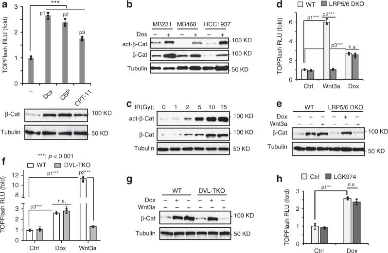
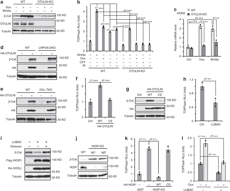
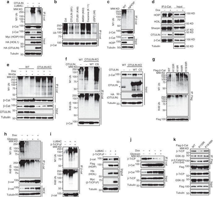
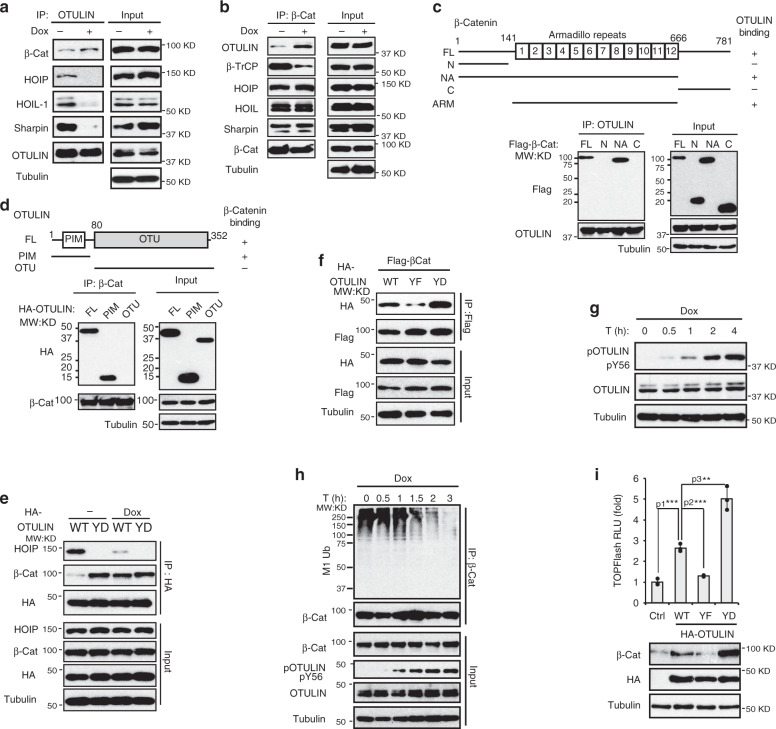
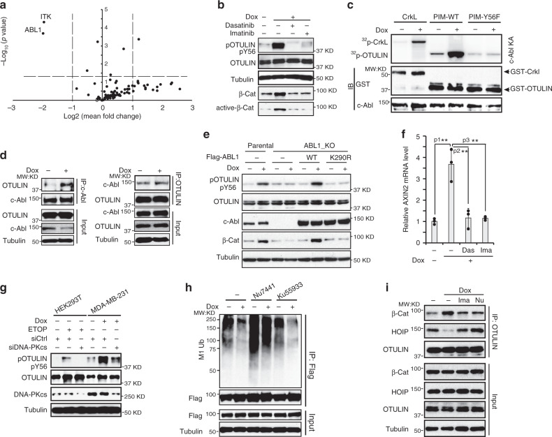
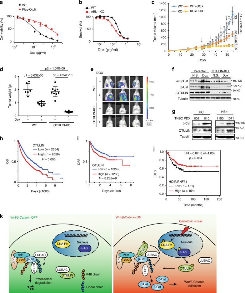
Dysregulated Wnt/β-catenin activation plays a critical role in cancer progression, metastasis, and drug resistance. Genotoxic agents such as radiation and chemotherapeutics have been shown to activate the Wnt/β-catenin signaling although the underlying mechanism remains incompletely understood. Here, we show that genotoxic agent-activated Wnt/β-catenin signaling is independent of the FZD/LRP heterodimeric receptors and Wnt ligands. OTULIN, a linear linkage-specific deubiquitinase, is essential for the DNA damage-induced β-catenin activation. OTULIN inhibits linear ubiquitination of β-catenin, which attenuates its Lys48-linked ubiquitination and proteasomal degradation upon DNA damage. The association with β-catenin is enhanced by OTULIN Tyr56 phosphorylation, which depends on genotoxic stress-activated ABL1/c-Abl. Inhibiting OTULIN or Wnt/β-catenin sensitizes triple-negative breast cancer xenograft tumors to chemotherapeutics and reduces metastasis. Increased OTULIN levels are associated with aggressive molecular subtypes and poor survival in breast cancer patients. Thus, OTULIN-mediated Wnt/β-catenin activation upon genotoxic treatments promotes drug resistance and metastasis in breast cancers.


Conflict of interest statement

The authors declare no competing interests.

Figures

Fig. 1
Fig. 1. DNA damage induces Wnt/β-catenin activation independent of canonical Wnt receptor complex FZD/LRP.
a TOPFlash assay and immunoblotting analysis of MDA-MB-231 cells treated with Dox (2 µg/ml), CBP (10 µg/ml), and CPT-11 (10 µM) for 24 h. n = 3 independent experiments. p1 = 2.25E−05, p2 = 8.53E−05, p3 = 0.000118. b Immunoblotting analysis of total and active-β-catenin in MDA-MB-231, MDA-MB-468, and HCC1937 cells treated with Dox (2 µg/ml) for 24 h. c Immunoblotting analysis of MDA-MB-231 cells exposed to IR with increasing doses. TOPFlash assay (d) and immunoblotting analysis of β-catenin expression (e) in parental and LRP5/6 DKO HEK293A cells after Wnt3a (20 ng/ml) and Dox (2 µg/ml) treatment for 24 h. n = 3 independent experiments in d. p1 = 9.93E−06, p2 = 1.09E−05, p3 = 1.39E−05. TOPFlash assay (f) and immunoblotting analysis of β-catenin expression (g) in parental and DVL1/2/3-TKO HEK293T cells treated as in d. n = 3 independent experiments in f. p1 = 2.95E−06, p2 = 3.21E−06, p3 = 0.000104. h TOPFlash assay of MDA-MB-231 cells treated with Dox (2 µg/ml) for 24 h, with or without pretreatment of porcupine inhibitor LGK974 (100 nM). n = 3 independent experiments. p1 = 0.000107. The statistical analysis in a, d, f and h was performed by two-sided unpaired t-test and p values are indicated as p < 0.05, ∗∗p < 0.01, and ∗∗∗p < 0.001. Data are presented as Mean ± SD. Source data are provided as a Source Data file.
Fig. 2
Fig. 2. OTULIN mediates genotoxic Wnt activation by inhibiting linear ubiquitination.
a Immunoblotting analysis of β-catenin in parental and OTULIN knockout MDA-MB-231 cells treated with Dox (2 µg/ml) or Wnt3a (20 ng/ml) for 24 h. b TOPFlash assay of parental and OTULIN knockout MDA-MB-231 cells treated with Wnt3a (20 ng/ml), Dox (2 µg/ml), CPT-11 (10 µM) or IR (10 Gy) for 24 h. n = 3 independent experiments. p1 = 4.39E−06, p2 = 3.55E−05, p3 = 1.56E−05, p4 = 5.48E−05, p5 = 8.73E−06, p6 = 5.22E−05, p7 = 1.59E−05, p8 = 1.86E−05, p9 = 3.29E−05. c qPCR analysis of AXIN2 mRNA level in parental and OTULIN knockout MDA-MB-231 cells treated with Dox (2 µg/ml) or Wnt3a (20 ng/ml) for 24 h. n = 3 independent experiments. p1 = 0.000188, p2 = 0.000503, p3 = 0.000117, p4 = 0.00413. d Immunoblotting analysis of β-catenin expression in parental and LRP5/6-DKO HEK293A cells with or without HA-OTULIN overexpression. e Immunoblotting analysis of β-catenin expression in parental and DVL1/2/3-TKO HEK293T cells with or without HA-OTULIN overexpression. TOPFlash assay (f) and immunoblotting analysis of β-catenin (g) in MDA-MB-231 cells transfected with HA-OTULIN wildtype (WT) or catalytic activity defect C129S mutant (CS). n = 3 independent experiments in f. p1 = 0.000783, p2 = 0.000497. h TOPFlash assay of HEK293T cells transfected with LUBAC components HOIP and HOIL. n = 3 independent experiments. p1 = 0.00670. i Immunoblotting analysis of β-catenin in MDA-MB-231 cells transfected with HOIP and HOIL and treated with or without LUBAC inhibitor Gliotoxin (1 µM) for 24 h. j Immunoblotting analysis of β-catenin expression in parental and HOIP knockout HEK293T cells. k TOPFlash assay of parental, HOIP knockout, or HOIP-KO HEK293T cells reconstituted with HOIP WT or C885S mutant. n = 3 independent experiments. p1 = 4.06E−05, p2 = 9.49E−06. l TOPFlash assay of MDA-MB-231 cells transfected with HOIP and HOIL and treated with Dox (2 µg/ml) for 24 h. n = 3 independent experiments. p1 = 0.000297, p2 = 0.000717, p3 = 0.000462. The statistical analysis in b, c, f, h, k and l was performed by two-sided unpaired t-test and p values are indicated as p < 0.05, ∗∗p < 0.01, and ∗∗∗p < 0.001. Data are presented as Mean±SD. Source data are provided as a Source Data file.
Fig. 3
Fig. 3. OTULIN inhibits linear ubiquitination of β-catenin.
a M1-linked ubiquitination of immunoprecipitated β-catenin from MDA-MB-231 cells transfected with LUBAC (HOIP + HOIL1) or along with HA-OTULIN. b UbiCREST analysis of β-catenin ubiquitination. MDA-MB-231 cells were treated with MG132 (10 µM) for 16 h. Then, β-Catenin was immunoprecipitated and incubated with indicated DUBs. The ubiquitination of β-Catenin precipitates after DUB digestion was analyzed by Western blot. c Linear ubiquitination of immunoprecipitated β-catenin from parental or HOIP knockout HEK293T cells. d Immunoblotting of co-immunoprecipitated proteins with β-Catenin in MDA-MB-231 cells treated with Dox (2 µg/ml) and MG132 (10 µM) for 8 h. e Linear ubiquitination of β-Catenin immunoprecipitates from parental and OTULIN knockout MDA-MB-231 cells treated as indicated. f Levels of linear Ub chain and K48 Ub chain in β-Catenin immunoprecipitates from parental, OTULIN knockout MDA-MB-231 cells or OTULIN-KO cells reconstituted with OTULIN-WT or –C129S mutant. *: endogenous OTULIN. g Linear and K48 ubiquitination of Flag-β-catenin WT, K133R, K19R, K49R, or K19/49R mutants in HEK293T cells. h Linear and K48 ubiquitination of β-Catenin immunoprecipitates in MDA-MB-231 cells treated with Dox (2 µg/ml), Gliotoxin (1 µM) or in combination for 24 h. i Linear and K48 ubiquitination of β-Catenin immunoprecipitates in HEK293T cells transfected with LUBAC and F-box-deleted β-TrCP mutant. j The analysis of β-catenin and β-TrCP interaction in MDA-MB-231 cells treated as in h. k Co-IP assay with anti-Flag antibody in HEK293T cells transfected with Flag-β-catenin WT, K133R, or K19/49 R. Source data are provided as a Source Data file.
Fig. 4
Fig. 4. OTULIN Tyr56 phosphorylation upon DNA damage promotes its association with β-catenin.
a Co-IP assay with anti-OTULIN antibody in MDA-MB-231 cells treated by Dox (2 µg/ml) and MG132 (10 µM) for 8 h. b Co-IP assay with anti-β-Catenin antibody in MDA-MB-231 cells treated as in a. c Mapping of β-Catenin domain required for interacting with OTULIN. d Mapping of OTULIN domain required for interacting with β-Catenin. e MDA-MB-231 cells transfected with OTULIN WT or Y56D mutant were treated with MG132 (10 µM) for 8 h, then with MG132 in combination of Dox (2 µg/ml) for 8 h. f The Co-IP analysis of Flag-β-Catenin and HA-OTULIN WT, Y56F or Y56D in HEK293T cells. MG132 (10 µM) was added to the culture 16 h before harvest. g Detection of OTULIN phosphorylation at Tyr56 in MDA-MB-231 cells treated with Dox (2 µg/ml) for times indicated. h Linear ubiquitination of precipitated β-Catenin from MDA-MB-231 cells treated with Dox (2 µg/ml) for times as indicated. i TOPFlash assay in MDA-MB-231 cells transfected with OTULIN WT, Y56F, or Y56D. n = 3 independent experiments. p1 = 0.000300, p2 = 0.000286, p3 = 0.00247. The statistical analysis in i was performed by two-sided unpaired t-test and p value is indicated as p < 0.05, ∗∗p < 0.01, and ∗∗∗p < 0.001. Data are presented as Mean ± SD. Source data are provided as a Source Data file.
Fig. 5
Fig. 5. DNA damage-activated c-Abl is required for OTULIN phosphorylation.
a Tyrosine kinase siRNA sublibrary was used to screen the kinase responsible for OTULIN Tyr56 phosphorylation. The pooled results from three replicates were presented as a volcano plot. n = 3 independent experiments. b Detection of OTULIN phosphorylation at Tyr56 in MDA-MB-231 cells pretreated with Dasatinib (10 µM) or Imatinib (10 µM) and subsequently treated with Dox (2 µg/ml) for 90 min. c c-Abl kinase assay in MDA-MB-231 cells treated with Dox (2 µg/ml) for 90 min, using recombinant CrkL, OTULIN-WT or -Y56F mutant PIM fragment as substrates. d Co-IP analysis of c-Abl and OTULIN in MDA-MB-231 cells treated with Dox (2 µg/ml) for 90 min. e Detection of OTULIN phosphorylation at Tyr56 in parental, ABL1 knockout MDA-MB-231 cells, and ABL1-KO cells transfected with ABL1-WT or K290R mutant after Dox (2 µg/ml) treatment for 90 min. f qPCR analysis of AXIN2 mRNA level in MDA-MB-231 cells pretreated with Dasatinib (10 µM) or Imatinib (10 µM) and subsequently treated with Dox (2 µg/ml) for 24 h. n = 3 independent experiments. Data are presented as Mean±SD, p1 = 0.00388, p2 = 0.00788, p3 = 0.00455. g Immunoblotting analysis of HEK293T and MDA-MB-231 cells, with or without DNA-PKcs knockdown, treated with Etoposide (10 µM) and Dox (2 µg/ml) for 90 min, respectively. h Linear ubiquitination in Flag-β-Catenin-immunoprecipitates from MDA-MB-231 cells pretreated with DNA-PK inhibitor Nu7441 (10 µM) or ATM inhibitor Ku55933 (10 µM) and subsequently treated with MG132 (10 µM) and Dox (2 µg/ml) for 8 h. i Co-IP analysis of OTULIN, β-Catenin and HOIP in MDA-MB-231 cells pretreated with Imatinib (10 µM) or Nu7441 (10 µM) and subsequently treated with MG132 (10 µM) and Dox (2 µg/ml) for 8 h. The statistical analysis in f was performed by two-sided unpaired t-test and p value is indicated as p < 0.05, ∗∗p < 0.01, and ∗∗∗p < 0.001. Source data are provided as a Source Data file.
Fig. 6
Fig. 6. Genotoxic Wnt activation promotes drug resistance and metastasis in breast cancer.
a Cell viability of parental and PyMT cells stably expressing OTULIN at 72 h after Dox treatment. Dox IC50 is 0.1 µg/ml and 0.75 µg/ml in parental and Flag-OTULIN PyMT cells, respectively. n = 3 independent experiments. b Cell viability of parental and ABL1 knockout MDA-MB-231 cells in response to Dox treatment for 72 h. Dox IC50 is 0.063 µg/ml and 0.016 µg/ml for parental and ABL1 knockout MDA-MB-231 cells, respectively. n = 3 independent experiments. cf Orthotopic xenograft model generated with parental or OTULIN-KO LM2 cells were treated with Dox (1.5 mg/kg/wk). Tumor growth curves were shown in c. p1 = 7.69E−05, p2 = 2.07E−09. At the endpoint, tumors were dissected and weighed as shown in d. p1 = 8.63E−05, p2 = 1.07E−09, p3 = 4.24E−10. Lung metastasis was monitored by bioluminescent imaging (e). Tumor samples were examined by Western blot for indicated proteins (f). n = 5 mice/group. g Immunoblotting analysis of β-Catenin and OTULIN expression in TNBC PDX cells, treatment naïve: HCI-002 and HBrt1150; chemorefractory: HCI-010 and HBrt1071. Overall survival (h) and disease-free survival (i) analysis in breast cancer patients from a combined dataset (TCGA-TARGET-GTEx). j Disease-free survival analysis in TNBC patients who received systemic chemotherapy stratified on the levels of HOIP (KM-Plot, two-sided, no adjustment for multiple comparisons). k Graphic summary of OTULIN-mediated genotoxic Wnt signaling pathway. The statistical analysis in c and d was performed by two-sided unpaired t-test and in hj was performed by the two-sided Log-rank test. p values are shown as p < 0.05, ∗∗p < 0.01, and ∗∗∗p < 0.001. Data are presented as Mean ± SD. Source data are provided as a Source Data file.

References

    1. Nusse R, Clevers H. Wnt/beta-Catenin signaling, disease, and emerging therapeutic modalities. Cell. 2017;169:985–999. - PubMed
    1. Stamos JL, Weis WI. The beta-catenin destruction complex. Cold Spring Harb. Perspect. Biol. 2013;5:a007898. - PMC - PubMed
    1. Hart M, et al. The F-box protein beta-TrCP associates with phosphorylated beta-catenin and regulates its activity in the cell. Curr. Biol. 1999;9:207–210. - PubMed
    1. Kitagawa M, et al. An F-box protein, FWD1, mediates ubiquitin-dependent proteolysis of beta-catenin. EMBO J. 1999;18:2401–2410. - PMC - PubMed
    1. Nusse R. Wnt signaling. Cold Spring Harb. Perspect. Biol. 2012;4:a011163. - PMC - PubMed

Publication types

MeSH terms