. 2020 Aug 7;10(1):13332.
doi: 10.1038/s41598-020-70258-w.

A versatile bulk electrotransfection protocol for murine embryonic fibroblasts and iPS cells

Affiliations

A versatile bulk electrotransfection protocol for murine embryonic fibroblasts and iPS cells

Shahin Eghbalsaied et al. Sci Rep. .

Abstract

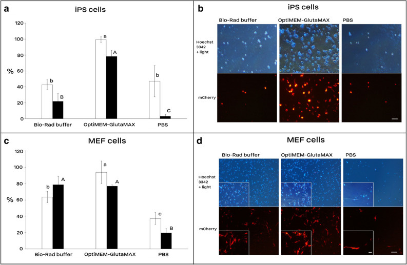
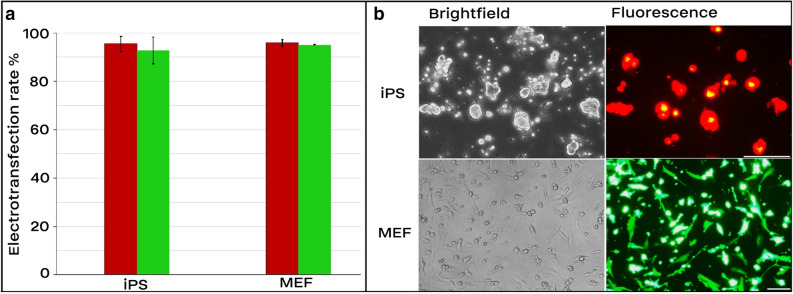
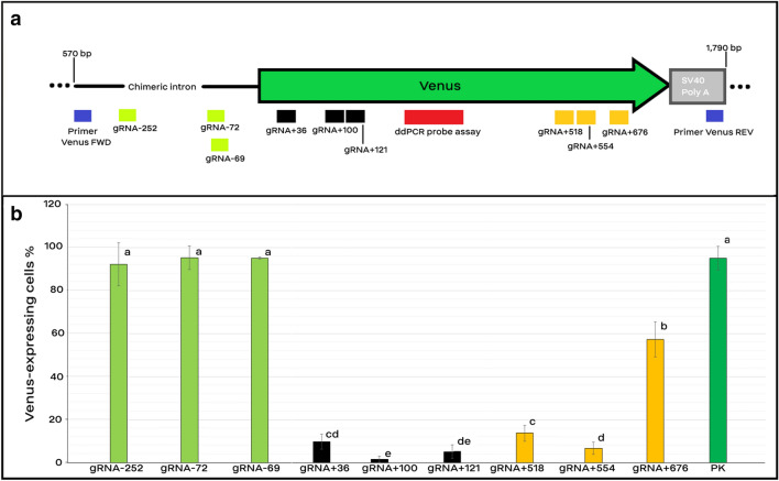
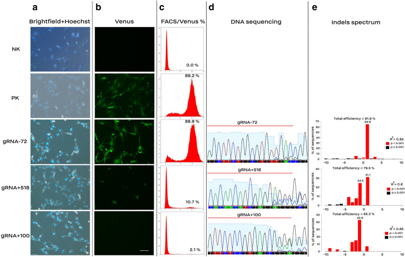
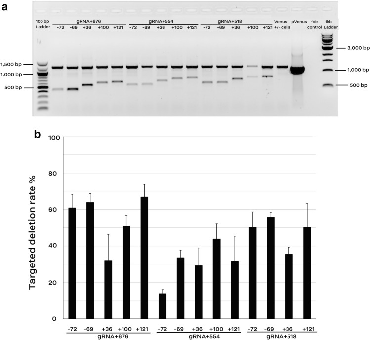
Although electroporation has been widely accepted as the main gene transfer tool, there is still considerable scope to improve the electroporation efficiency of exogenous DNAs into primary cells. Here, we developed a square-wave pulsing protocol using OptiMEM-GlutaMAX for highly efficient transfection of murine embryonic fibroblasts (MEF) and induced pluripotency stem (iPS) cells using reporter genes as well as gRNA/Cas9-encoding plasmids. An electrotransfection efficiency of > 95% was achieved for both MEF and iPS cells using reporter-encoding plasmids. The protocol was efficient for plasmid sizes ranging from 6.2 to 13.5 kb. Inducing the error prone non-homologous end joining repair by gRNA/Cas9 plasmid transfection, a high rate of targeted gene knockouts of up to 98% was produced in transgenic cells carrying a single-copy of Venus reporter. Targeted deletions in the Venus transgene were efficiently (up to 67% deletion rate) performed by co-electroporation of two gRNA-encoding plasmids. We introduced a plasmid electrotransfection protocol which is straight-forward, cost-effective, and efficient for CRISPRing murine primary cells. This protocol is promising to make targeted genetic engineering using the CRISPR/Cas9 plasmid system.


Conflict of interest statement

The authors declare no competing interests.

Figures

Figure 1
Figure 1
Cell electrotransfection and viability rates using Bio-Rad buffer, OptiMEM-GlutaMAX, and PBS. Electrotransfection efficiency of mouse iPS (a and b) and MEF cells (c and d). In each electroporation reaction, 20 µg (1.5–2.5 µg/µl) of the reporter plasmid (encoding mCherry) was pre-mixed with cells and underwent electroporation using the square-wave protocol consisted of 250 V for iPS and 300 V for MEF cells, 2 pulses, each 10 ms length, 10 s interval, and 4 mm cuvette. The reporter expression was assessed 36 h after electroporation under a fluorescence microscope. White and black bars are electrotransfection efficiency and cell viability, respectively. Bars with different A, B, C or a, b, c letters are significantly different (p value < 0.05). Scale bar is 100 µm; for MEF cells, a higher magnified photo is indented in the left part of each image with a scale bar of 5 µm. Results are means and standard deviation (n > 3).
Figure 2
Figure 2
Electrotransfection efficiency of MEF and iPS cells. In each electroporation reaction, 20 µg (1.5–2.5 µg/µl) of either pT2-Venus and pT2-mCherry which encode Venus and mCherry proteins, respectively, were pre-mixed with cells and underwent electroporation. The following electroporation program was used: square-wave protocol with either 250 V for iPS or 300 V for MEF cells, each 10 ms pulse length, 2 pulses, 10 s pulse interval, and 4 mm cuvette. (a) Transfection efficiency. Transfected cells for mCherry and Venus are depicted by red and green bars, respectively. (b) Expression of Venus and mCherry 36 h after electroporation. Scale bar for iPS cells equals 100 µm. Scale Bar in fibroblasts is 10 µm. Results are means and standard deviation (n > 3).
Figure 3
Figure 3
Target site and knockout efficiency of gRNAs. (a) Schematic presentation of gRNA target location on the Venus transgene. Nine gRNAs were designed to target the promoter region (− 252, − 72, and − 69), 5′ region (36, 100, and 121), and 3′ region (518, 554, and 676) of Venus transgene which are depicted with white-green, black, and yellow bars, respectively. The target site of the digital PCR assay is depicted in a red box. (b) Efficiency of different gRNAs for making Venus knockout. Cells were treated with puromycin and were screened for the Venus transgene under a fluorescence microscope 10 days after the electroporation (n > 3). Bars with common letters are not significantly different (p-value < 0.05). Cells with one copy of Venus, which did not undergo any electroporation treatment, were considered as the positive control for the Venus expression (PK).
Figure 4
Figure 4
Knockout of Venus transgene by inducing indels using gRNA-encoding plasmids. MEF cells carrying a single-copy of the Venus transgene were electrotransfected with different gRNA-encoding plasmids (9.1 kb length). Induction of indels using gRNA-72 had no effect on the Venus expression, whereas gRNA + 100 resulted in loss of the Venus signal. Electroporated cells were selected against puromycin and screened 10 days after the electroporation. (a and b) Cells were stained with Hoechst 33342 and the efficiency of Venus knockout was assessed. Scale bar equals 10 µm. (c) Histoplots of the FACS results. (d) Partial sequencing results of the amplified Venus transgene. Target sites of the respective gRNAs are indicated by a red line, the first three nucleotides of the electropherogram are cropped to fit the image into the column. (e) Indels spectrum calculated by online tool for TIDE analysis. Cells with no copy of Venus were considered as the negative control (NK), and cells carrying a copy of Venus, without any electroporation treatment, were considered as the positive control for the Venus expression (PK). The following electroporation program was used: square-wave protocol: 20 µg plasmid (1.5–2.5 µg/µl), 300 V, 2 pulses, each 10 ms pulse length, 10 s pulse interval, and 4 mm cuvette (n > 3).
Figure 5
Figure 5
Targeted deletions of Venus transgene by co-electroporation of two gRNA-encoding plasmids. (a) End-point PCR showed deletion of 15 different fragments from the original amplicon (1204 bp) using pairwise combination of gRNAs targeting the promoter/ 5′ region (− 72, − 69, + 36, + 100, and + 121) and the 3′ region (+ 518, + 554, and + 676) of Venus cDNA. The primer set (Venus-Forward1 and Venus-Reverse2) amplified both the shortened and original fragments. Genomic DNA from MEF cells carrying no-copy of Venus was considered as the negative controls (− Ve control). (b) Digital-PCR results for quantification of the rate of targeted deletion via a primer–probe assay. The primer–probe assay was designed to amplify only the wild type fragment. The deletion rate was calculated as the rate of copy number/µl in the co-electroporated groups divided by that of their control (non-treated) counterparts. Because of a poor DNA quality, the combined group of gRNA + 518 and + 100 was removed from the digital PCR analysis. Data are depicted as average ± standard error (n > 3).

Similar articles

Cited by

References

    1. Sakuma T, Nishikawa A, Kume S, Chayama K, Yamamoto T. Multiplex genome engineering in human cells using all-in-one CRISPR/Cas9 vector system. Sci. Rep. 2014;4:5400. doi: 10.1038/srep05400. - DOI - PMC - PubMed
    1. Han X, et al. CRISPR-Cas9 delivery to hard-to-transfect cells via membrane deformation. Sci. Adv. 2015;1:e1500454. doi: 10.1126/sciadv.1500454. - DOI - PMC - PubMed
    1. Liu C, Zhang L, Liu H, Cheng K. Delivery strategies of the CRISPR-Cas9 gene-editing system for therapeutic applications. J. Control. Release. 2017;266:17–26. doi: 10.1016/j.jconrel.2017.09.012. - DOI - PMC - PubMed
    1. Gresch O, et al. New non-viral method for gene transfer into primary cells. Methods. 2004;33:151–163. doi: 10.1016/j.ymeth.2003.11.009. - DOI - PubMed
    1. Weaver JC, Smith KC, Esser AT, Son RS, Gowrishankar T. A brief overview of electroporation pulse strength–duration space: A region where additional intracellular effects are expected. Bioelectrochemistry. 2012;87:236–243. doi: 10.1016/j.bioelechem.2012.02.007. - DOI - PMC - PubMed

Publication types

MeSH terms

Substances