Unique inducible filamentous motility identified in pathogenic Bacillus cereus group species
- PMID: 32770116
- PMCID: PMC7784679
- DOI: 10.1038/s41396-020-0728-x
Unique inducible filamentous motility identified in pathogenic Bacillus cereus group species
Abstract
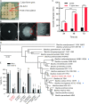
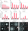
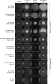
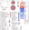
Active migration across semi-solid surfaces is important for bacterial success by facilitating colonization of unoccupied niches and is often associated with altered virulence and antibiotic resistance profiles. We isolated an atmospheric contaminant, subsequently identified as a new strain of Bacillus mobilis, which showed a unique, robust, rapid, and inducible filamentous surface motility. This flagella-independent migration was characterized by formation of elongated cells at the expanding edge and was induced when cells were inoculated onto lawns of metabolically inactive Campylobacter jejuni cells, autoclaved bacterial biomass, adsorbed milk, and adsorbed blood atop hard agar plates. Phosphatidylcholine (PC), bacterial membrane components, and sterile human fecal extracts were also sufficient to induce filamentous expansion. Screening of eight other Bacillus spp. showed that filamentous motility was conserved amongst B. cereus group species to varying degrees. RNA-Seq of elongated expanding cells collected from adsorbed milk and PC lawns versus control rod-shaped cells revealed dysregulation of genes involved in metabolism and membrane transport, sporulation, quorum sensing, antibiotic synthesis, and virulence (e.g., hblA/B/C/D and plcR). These findings characterize the robustness and ecological significance of filamentous surface motility in B. cereus group species and lay the foundation for understanding the biological role it may play during environment and host colonization.
Conflict of interest statement
The authors declare that they have no conflict of interest.
Figures


Similar articles
-
Salt-induced Reduction of Hyperswarming Motility in Bacillus cereus MHS is Associated with Reduction in Flagellation, Nanotube Formation and Quorum Sensing Regulator plcR.Curr Microbiol. 2025 May 28;82(7):313. doi: 10.1007/s00284-025-04288-w. Curr Microbiol. 2025. PMID: 40437124
-
PlcR is a pleiotropic regulator of extracellular virulence factor gene expression in Bacillus thuringiensis.Mol Microbiol. 1999 Jun;32(5):1043-53. doi: 10.1046/j.1365-2958.1999.01419.x. Mol Microbiol. 1999. PMID: 10361306
-
FlhF, a signal recognition particle-like GTPase, is involved in the regulation of flagellar arrangement, motility behaviour and protein secretion in Bacillus cereus.Microbiology (Reading). 2007 Aug;153(Pt 8):2541-2552. doi: 10.1099/mic.0.2006/005553-0. Microbiology (Reading). 2007. PMID: 17660418
-
Bacillus cereus ATCC 14579 RpoN (Sigma 54) Is a Pleiotropic Regulator of Growth, Carbohydrate Metabolism, Motility, Biofilm Formation and Toxin Production.PLoS One. 2015 Aug 4;10(8):e0134872. doi: 10.1371/journal.pone.0134872. eCollection 2015. PLoS One. 2015. PMID: 26241851 Free PMC article.
-
What sets Bacillus anthracis apart from other Bacillus species?Annu Rev Microbiol. 2009;63:451-76. doi: 10.1146/annurev.micro.091208.073255. Annu Rev Microbiol. 2009. PMID: 19514852 Review.
Cited by
-
Filamentous morphology of bacterial pathogens: regulatory factors and control strategies.Appl Microbiol Biotechnol. 2022 Sep;106(18):5835-5862. doi: 10.1007/s00253-022-12128-1. Epub 2022 Aug 22. Appl Microbiol Biotechnol. 2022. PMID: 35989330 Review.
-
Metabolomic profiling of VOC-driven interactions between Priestia megaterium and Bacillus licheniformis in a simulated rhizosphere using split petri dishes.Arch Microbiol. 2025 Aug 12;207(9):224. doi: 10.1007/s00203-025-04426-9. Arch Microbiol. 2025. PMID: 40794197 Free PMC article.
-
Methionine Sulfoxide Reductases Contribute to Anaerobic Fermentative Metabolism in Bacillus cereus.Antioxidants (Basel). 2021 May 20;10(5):819. doi: 10.3390/antiox10050819. Antioxidants (Basel). 2021. PMID: 34065610 Free PMC article.
-
Bacillus Species as Direct-Fed Microbial Antibiotic Alternatives for Monogastric Production.Probiotics Antimicrob Proteins. 2023 Feb;15(1):1-16. doi: 10.1007/s12602-022-09909-5. Epub 2022 Jan 29. Probiotics Antimicrob Proteins. 2023. PMID: 35092567 Free PMC article. Review.
-
Sources, transmission, and tracking of sporeforming bacterial contaminants in dairy systems.JDS Commun. 2023 Nov 17;5(2):172-177. doi: 10.3168/jdsc.2023-0428. eCollection 2024 Mar. JDS Commun. 2023. PMID: 38482119 Free PMC article. Review.
References
-
- Ben-Jacob E, Finkelshtein A, Ariel G, Ingham C. Multispecies swarms of social microorganisms as moving ecosystems. Trends Microbiol. 2016;24:257–69.. - PubMed
Publication types
MeSH terms
Substances
Supplementary concepts
Grants and funding
LinkOut - more resources
Full Text Sources
Molecular Biology Databases
Miscellaneous

