. 2020 Aug 8;10(1):139.
doi: 10.1186/s13568-020-01073-9.

Surface display of p75, a Lactobacillus rhamnosus GG derived protein, on Bacillus subtilis spores and its antibacterial activity against Listeria monocytogenes

Affiliations

Surface display of p75, a Lactobacillus rhamnosus GG derived protein, on Bacillus subtilis spores and its antibacterial activity against Listeria monocytogenes

Soo Ji Kang et al. AMB Express. .

Abstract

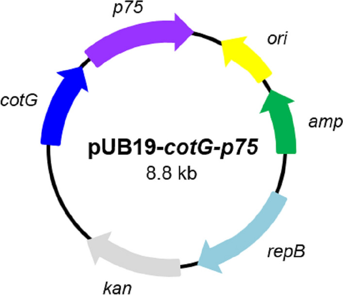
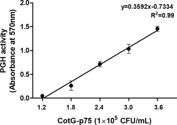
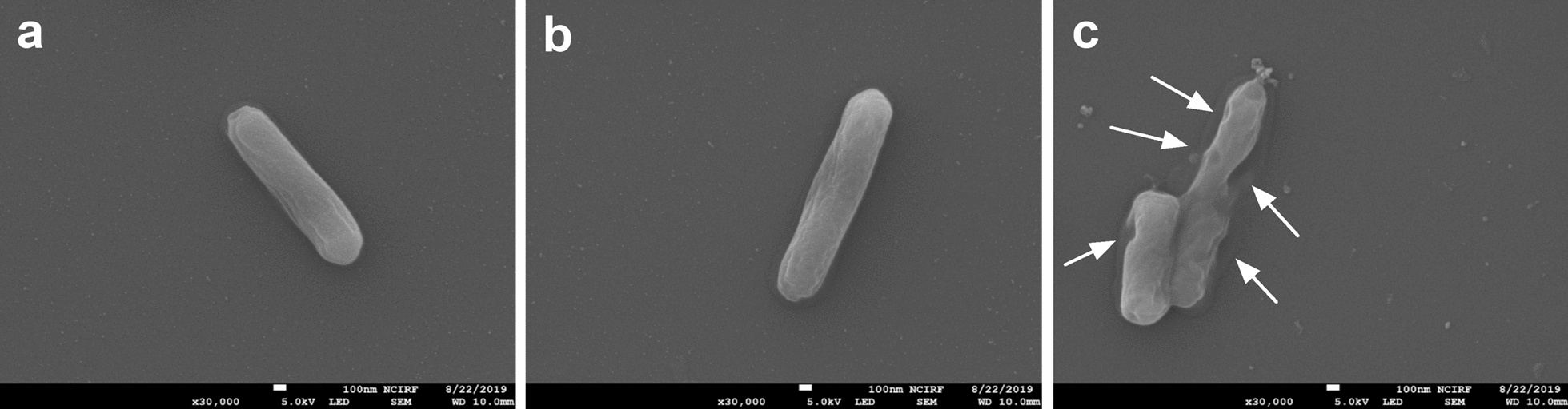
Lactobacillus rhamnosus p75 protein with peptidoglycan hydrolase (PGH) activity is one of the key molecules exhibiting anti-apoptotic and cell-protective activity for human intestinal epithelial cells. In this study, with the goal of developing new probiotics, the p75 protein was displayed on the surface of Bacillus subtilis spores using spore coat protein CotG as an anchoring motif. The PGH activity, stability, and the antibacterial activity of the spore-displayed p75 (CotG-p75) protein were also investigated. The PGH activity of the CotG-p75 against peptidoglycan extracted from B. subtilis was confirmed by the ninhydrin test. Under various harsh conditions, compared to the control groups, the PGH activities of CotG-p75 were very stable in the range of pH 3-7 and maintained at 70% at 50 °C. In addition, the antibacterial activity of CotG-p75 against Listeria monocytogenes was evaluated by a time-kill assay. After 6 h incubation in phosphate-buffered saline, CotG-p75 reduced the number of viable cells of L. monocytogenes by up to 2.0 log. Scanning electron microscopy analysis showed that the cell wall of L. monocytogenes was partially damaged by the treatment with CotG-p75. Our preliminary results show that CotG-p75 could be a good candidate for further research to develop new genetically engineered probiotics.

Keywords: Antibacterial activity; L. monocytogenes; Spore surface display; p75 protein.


Conflict of interest statement

The authors declare that they have no competing interests.

Figures

Fig. 1
Fig. 1
Plasmid map of the recombinant plasmid pUB19-cotG-p75. The ori and repB represent the replication origin and replication protein B, respectively. The amp and kan represent ampicillin and kanamycin resistance markers, respectively. The cotG and p75 represent the spore coat protein CotG encoding gene of B. subtilis and the p75 protein encoding gene of L. rhamnosus GG, respectively
Fig. 2
Fig. 2
Determination of the peptidoglycan hydrolase (PGH) activity of CotG-p75 measured by the ninhydrin method. After treatment of peptidoglycan with different concentrations of CotG-p75 at 37 °C for 15 min, the absorbance of each sample was measured at 570 nm. All tests were performed in triplicate, and the data are presented as mean ± standard deviation
Fig. 3
Fig. 3
Relative peptidoglycan hydrolase activity of CotG-p75 after heat (a) and pH (b) treatments. Relative activity was calculated by defining its activity at 25 °C and pH 7 as 100%. All tests were performed in triplicate, and the data are presented as mean ± standard deviation. Statistical analysis was performed by an unpaired two-tailed t-test. Asterisks indicate a significance difference from the control (*p < 0.05, **p < 0.01, ***p < 0.001)
Fig. 4
Fig. 4
Antibacterial effect of CotG-p75 against L. monocytogenes. Cells were treated with different concentrations of CotG-p75 in PBS at 37 °C. The control groups were treated with no spores (closed square) or wild-type spores (closed triangle). The number of viable cells was measured at intervals of 1 h during the 6 h incubation. All tests were performed in triplicate, and the data are presented as mean ± standard deviation. Statistical analysis was performed by an unpaired two-tailed t-test
Fig. 5
Fig. 5
SEM images of L. monocytogenes untreated (a) and treated with wild type spores (b), and CotG-p75 (c) in PBS for 6 h at 37 °C. Arrows indicate damaged cell surfaces

References

    1. Anagnostopoulos C, Crawford IP. Transformation studies on the linkage of markers in the tryptophan pathway in Bacillus subtilis. PNAS. 1961;47:378–390. - PMC - PubMed
    1. Atrih A, Bacher G, Allmaier G, Williamson MP, Foster SJ. Analysis of peptidoglycan structure from vegetative cells of Bacillus subtilis 168 and role of PBP 5 in peptidoglycan maturation. J Bacteriol. 1999;181(3):3956–3966. - PMC - PubMed
    1. Baba T, Schneewind O. Target cell specificity of a bacteriocin molecule: a C-terminal signal directs lysostaphin to the cell wall of Staphylococcus aureus. EMBO J. 1996;15(18):4789. - PMC - PubMed
    1. Barry AL, Craig WC, Nadler H, Reller LB, Sanders CC, Swenson JM (1999) Methods for determining bactericidal activity of antimicrobial agents; approved guideline. NCCLS document M26-A. National Committee for Clinical Laboratory Standards, Wayne, Pennsylvania
    1. Bäuerl C, Perez-Martinez G, Yan F, Polk DB, Monedero V. Functional analysis of the p40 and p75 proteins from Lactobacillus casei BL23. J Mol Microb Biotech. 2010;19:231–241. - PMC - PubMed