. 2020 Aug 8;21(1):208.
doi: 10.1186/s12931-020-01468-z.

Functional glutamate transporters are expressed in the carotid chemoreceptor

Affiliations

Functional glutamate transporters are expressed in the carotid chemoreceptor

Chaohong Li et al. Respir Res. .

Abstract

Background: The carotid body (CB) plays a critical role in cyclic intermittent hypoxia (CIH)-induced chemosensitivity; however, the underlying mechanism remains uncertain. We have demonstrated the presence of multiple inotropic glutamate receptors (iGluRs) in CB, and that CIH exposure alters the level of some iGluRs in CB. This result implicates glutamatergic signaling in the CB response to hypoxia. The glutamatergic neurotransmission is not only dependent on glutamate and glutamate receptors, but is also dependent on glutamate transporters, including vesicular glutamate transporters (VGluTs) and excitatory amino acid transporters (EAATs). Here, we have further assessed the expression and distribution of VGluTs and EAATs in human and rat CB and the effect of CIH exposure on glutamate transporters expression.

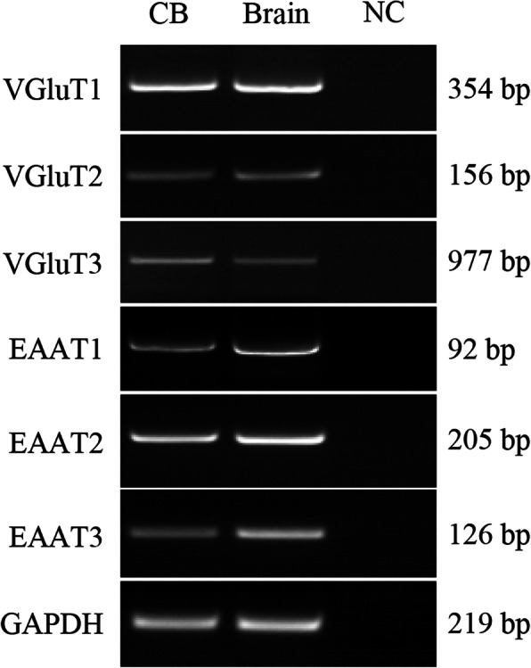
Methods: The mRNA of VGluTs and EAATs in the human CB were detected by RT-PCR. The protein expression of VGluTs and EAATs in the human and rat CB were detected by Western blot. The distribution of VGluT3, EAAT2 and EAAT3 were observed by immunohistochemistry staining and immunofluorescence staining. Male Sprague-Dawley (SD) rats were exposed to CIH (FIO2 10-21%, 3 min/3 min for 8 h per day) for 2 weeks. The unpaired Student's t-test was performed.

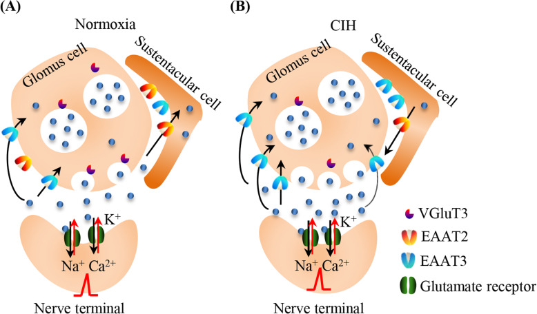
Results: Here, we report on the presence of mRNAs for VGluT1-3 and EAAT1-3 in human CB, which is consistent with our previous results in rat CB. The proteins of VGluT1 and 3, EAAT2 and 3, but not VGluT2 and EAAT1, were detected with diverse levels in human and rat CB. Immunostaining showed that VGluT3, the major type of VGluTs in CB, was co-localized with tyrosine hydroxylase (TH) in type I cells. EAAT2 and EAAT3 were distributed not only in type I cells, but also in glial fibrillary acidic protein (GFAP) positive type II cells. Moreover, we found that exposure of SD rats to CIH enhanced the protein level of EAAT3 as well as TH, but attenuated the levels of VGluT3 and EAAT2 in CB.

Conclusions: Our study suggests that glutamate transporters are expressed in the CB, and that glutamate transporters may contribute to glutamatergic signaling-dependent carotid chemoreflex to CIH.

Keywords: Carotid body; Cyclic intermittent hypoxia; Excitatory amino acid transporter; Glutamate; Vesicular glutamate transporter.


Conflict of interest statement

The authors declare that they have no competing interests.

Figures

Fig. 1
Fig. 1
Expression of VGluTs and EAATs mRNAs in human CB. RT-PCR showed expression of mRNAs of VGluTs and EAATs in human CB (CB, left lane). RNA extracted from human brain cerebral cortex was used as positive control (Brain, middle lane). The DEPC-H2O, instead of RNA, was used in RT reaction to obtain a negative cDNA control. An equal volume of the negative cDNA control was used in the PCR as PCR negative control (NC, right lane). VGluT: vesicular glutamate transporter; EAAT: excitatory amino acid transporter
Fig. 2
Fig. 2
Expression of VGluTs and EAATs proteins in human and rat CB. Western blot showed expression of proteins of VGluTs and of EAATs in rat and human CB (CB, left lane). Protein extracted from human and rat brain cerebral cortex was used as positive control (Brain, right lane). VGluT: vesicular glutamate transporter; EAAT: excitatory amino acid transporter
Fig. 3
Fig. 3
Immunohistochemical staining of VGluT3, EAAT2 and EAAT3 in the rat CB. Immunohistochemical staining showing the location of VGluT3 (A1–2), EAAT2 (B1–2) and EAAT3 (C1–2) in rat CB. D1-D2 are negative staining control obtained by omitting the primary antibody. A2-D2 are higher magnifications images of rectangle areas in A1-D1, respectively. The brown staining represents the glutamate transporter, while the blue staining represents hematoxylin-stained cell nuclei. VGluT: vesicular glutamate transporter; EAAT: excitatory amino acid transporter. Scale Bar = 50 μm for all images
Fig. 4
Fig. 4
Cellular distribution of VGluT3, EAAT2 and EAAT3 in the rat CB. A Double immunofluorescence staining of VGluT3 (red) and tyrosine hydroxylase (TH, green) in rat CB (a-c1). B Double immunofluorescence staining of VGluT3 (red) and glial fibrillary acidic protein (GFAP, green) in rat CB (d-f1). C Double immunofluorescence staining of EAAT2 (green) and TH (red) in rat CB (g-i1). D Double immunofluorescence staining of EAAT2 (green) and GFAP (red) in rat CB (j-l1). E Double immunofluorescence staining of EAAT3 (green) and TH (red) in rat CB (m-o1). F Double immunofluorescence staining of EAAT3 (green) and GFAP (red) in rat CB (p-r1). a1-r1 are higher magnifications images of rectangle areas in a-c, respectively. TH: the CB type I cells marker; GFAP: the CB type II cells marker; VGluT: vesicular glutamate transporter; EAAT: excitatory amino acid transporter. Scale bar = 25 μm for all images
Fig. 5
Fig. 5
CIH treatment increases the EAAT3 protein level and decreases the VGluT3 and EAAT2 protein level in the rat CB. a Western blot showed protein expression level of VGluT3, EAAT2 and EAAT3 in the rat CB of Con and CIH group rats. b Statistical analysis of VGluT3, EAAT2 and EAAT3 protein level in the rat CB after CIH treatment. The data were presented as means ± S.D. The standard deviation bars represent the standard deviation from 3 technical replicates. n = 8 in each group, *P < 0.05, **P < 0.01
Fig. 6
Fig. 6
Schematic representation of glutamatergic neurotransmission in the CB. a. Under normoxia, glutamate released from CB pre-synaptic type I cell acts on glutamate ionotropic receptors on its corresponding post-synaptic type I cell. The influx of Na+ and Ca2+ ions cause depolarization and generation of action potential. b. CIH causes CB neuronal damages, then accumulation of glutamate induced neurotoxicity, leading compensatory reduction of VGluT2 and EAAT2 as well as an increase of EAAT3

Similar articles

Cited by

References

    1. Nieto FJ, Young TB, Lind BK, Shahar E, Samet JM, Redline S, D'Agostino RB, Newman AB, Lebowitz MD, Pickering TG. Association of sleep-disordered breathing, sleep apnea, and hypertension in a large community-based study. Sleep Heart Health Study JAMA. 2000;283(14):1829–1836. - PubMed
    1. Sjöström C, Lindberg E, Elmasry A, Hägg A, Svärdsudd K, Janson C. Prevalence of sleep apnoea and snoring in hypertensive men: a population based study. Thorax. 2002;57(7):602–607. - PMC - PubMed
    1. Drager LF, Genta PR, Pedrosa RP, Nerbass FB, Gonzaga CC, Krieger EM, Lorenzi-Filho G. Characteristics and predictors of obstructive sleep apnea in patients with systemic hypertension. Am J Cardiol. 2010;105(8):1135–1139. - PubMed
    1. Fletcher EC, Lesske J, Qian W, Miller CC, 3rd, Unger T. Repetitive, episodic hypoxia causes diurnal elevation of systemic blood pressure in rats. Hypertension. 1992;19(6 Pt 1):555–561. - PubMed
    1. Fletcher EC, Lesske J, Culman J, Miller CC, Unger T. Sympathetic denervation blocks blood pressure elevation in episodic hypoxia. Hypertension. 1992;20(5):612–619. - PubMed

MeSH terms

Substances

LinkOut - more resources