. 2020 Aug 8;8(1):131.
doi: 10.1186/s40478-020-01009-1.

Demonstrating a reduced capacity for removal of fluid from cerebral white matter and hypoxia in areas of white matter hyperintensity associated with age and dementia

Affiliations

Demonstrating a reduced capacity for removal of fluid from cerebral white matter and hypoxia in areas of white matter hyperintensity associated with age and dementia

Matthew MacGregor Sharp et al. Acta Neuropathol Commun. .

Abstract

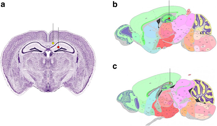
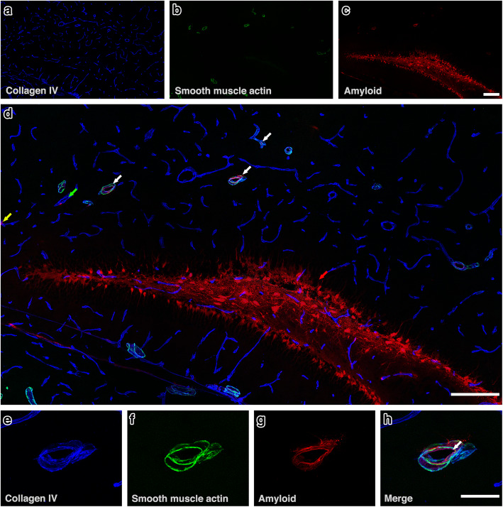
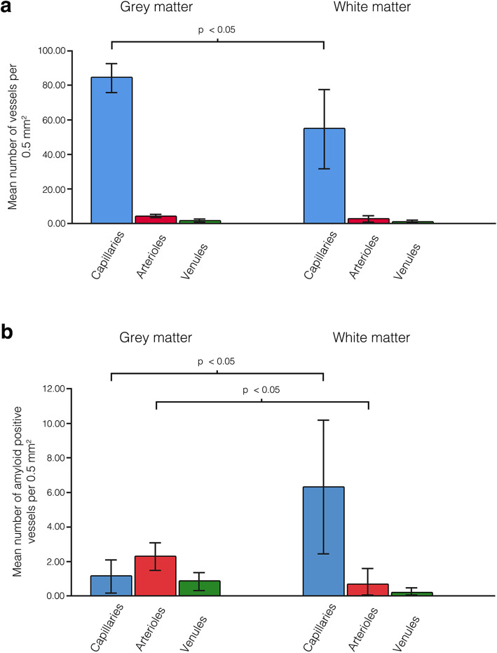
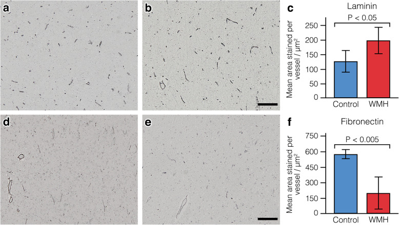
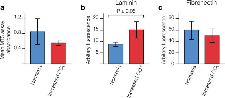
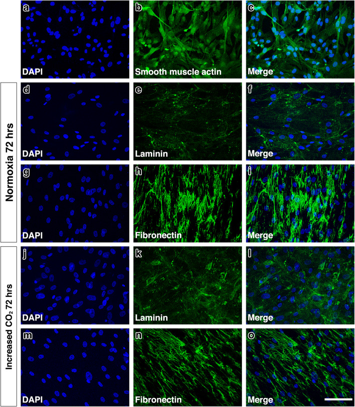
White matter hyperintensities (WMH) occur in association with dementia but the aetiology is unclear. Here we test the hypothesis that there is a combination of impaired elimination of interstitial fluid from the white matter together with a degree of hypoxia in WMH. One of the mechanisms for the elimination of amyloid-β (Aβ) from the brain is along the basement membranes in the walls of capillaries and arteries (Intramural Peri-Arterial Drainage - IPAD). We compared the dynamics of IPAD in the grey matter of the hippocampus and in the white matter of the corpus callosum in 10 week old C57/B16 mice by injecting soluble Aβ as a tracer. The dynamics of IPAD in the white matter were significantly slower compared with the grey matter and this was associated with a lower density of capillaries in the white matter. Exposing cultures of smooth muscle cells to hypercapnia as a model of cerebral hypoperfusion resulted in a reduction in fibronectin and an increase in laminin in the extracellular matrix. Similar changes were detected in the white matter in human WMH suggesting that hypercapnia/hypoxia may play a role in WMH. Employing therapies to enhance both IPAD and blood flow in the white matter may reduce WMH in patients with dementia.

Keywords: Fibronectin; Intramural periarterial drainage; Laminin; White matter hyperintensities.


Conflict of interest statement

The authors declare no competing interests.

Figures

Fig. 1
Fig. 1
Stereotaxic Injection sites. Injections were performed Anterior-Posterior − 2 mm from the Bregma represented by the red dot (hippocampus) and yellow dot (corpus callosum) in (a). Sagittal slices show the injection site for the hippocampus (Anterior-Posterior - 2 mm; Medial-Lateral 1.5 mm; Dorsal-Ventral 1.7 mm), red dot in (b) and corpus callosum (Anterior-Posterior -2 mm; Medial-Lateral 0.5 mm; Dorsal-Ventral - 1.3 mm), yellow dot in (c). Images adapted from www.mouse.brain-map.org
Fig. 2
Fig. 2
IPAD in hippocampus. (a -c) The distribution of amyloid-β in relation to collagen IV and smooth muscle actin at 7 min after intrahippocampal injection. c & d) Amyloid-β (red) was observed diffusely distributed in the parenchyma and co-localised (pink colour) with collagen IV in the walls of arterioles (white arrows), capillaries (yellow arrow) and few venules (green arrow). Representative high power image of an arteriole (e-h) shows amyloid-β (red) (g-h) in the wall of the blood vessel, indicated by the white arrow in (h). Scale bars a-d = 200 μm, e – h = 10 μm
Fig. 3
Fig. 3
IPAD in corpus callosum. (a-c) Distribution of amyloid-β in relation to collagen IV and smooth muscle actin. Amyloid-β (red) was observed along the white matter tracts (c & d) and co-localising (Pink) with collagen IV in the walls of capillaries (yellow arrows) and arterioles (white arrows). (e-h) An arteriole in the white matter - see box in (d) -showing Aβ in the tunica media. Merging of the blue collagen IV staining in the basement membranes in (e) with red amyloid (g) produces a pink colour of co-localisation in (h). (i-l) A leptomeningeal artery in the hippocampal fissure abutting on to the white matter shows Aβ co-localized (pink) with collagen IV in the tunica media (lower arrow) and in the adventitia (upper arrow). (m-p) shows red Aβ (arrow) in the wall of a white matter capillary that is also stained blue for collagen IV in the basement membrane. Scale bars a-d = 200 μm, e – h = 20 μm & i –p = 10 μm
Fig. 4
Fig. 4
Comparison of vessel density (a) and the density of vessels with fluorescent Aβ in their walls (b) between the hippocampus (grey matter) and corpus callosum (white matter). The density of arterioles with fluorescent Aβ in their vessel walls was significantly higher in the hippocampus but the density of capillaries with fluorescent Aβ in their vessel walls significantly higher in the corpus callosum (b) despite the significant reduction in density of capillaries in the corpus callosum (a). Error bars: +/− 2 SE
Fig. 5
Fig. 5
Laminin and fibronectin expression in the white matter in subjects with white matter hyperintensities and controls. a Representative image of laminin staining in the control group. b Representative image of laminin staining in the WMH group. c Histogram showing the mean area of laminin staining per vessel. d Representative image of fibronectin staining in the control group. e Representative image of fibronectin staining in the WMH group. f Histogram showing the mean area of fibronectin staining per vessel. Scale bars indicate 200um. Error bars indicate +/− 2 SE
Fig. 6
Fig. 6
Mean MTS absorbance and mean fluorescence intensity of HBVSMC exposed to increased levels of CO2. a Mean MTS absorbance was reduced but not significantly.b Laminin staining was significantly higher in cultures exposed to increased levels of CO2 compared to the normoxia cultures (p ≤ 0.05). c There was a trend towards a decrease in the fluorescence intensity of fibronectin staining but this was not statistically significant. Error bars: +/− 1 SE
Fig. 7
Fig. 7
Laminin and fibronectin staining in HBVSMC cultures after 72 h exposure to normoxia and hypoxia. Reference images (a – c) show the presence of smooth muscle actin in the HBVSMCs used in this study. The pattern of laminin staining (e,k) was similar in the HBVSMC cultures but there was a significant increase in expression with increased levels of CO2(l) compared to normoxice cultures (f). Fibronectin staining (h,n) had a web-like appearance in both cultures but staining appeared more intense in the normoxic cultures (i) compared to cultures exposed to increased levels of CO2(o). Cell density, indicated by DAPI stained nuclei (d, g, j, m), did not appear to be altered in normoxic conditions or conditions of increased levels of CO2. Scale bar = 50 μm
Fig. 8
Fig. 8
Aetiology of White Matter Hyperintensities (WMH) in the human brain. Two of the major functions of cerebral arteries, arterioles and capillaries are (a) Blood Flow and the supply of nutrients to brain tissues (Red Arrow) and (b) Elimination of Interstitial Fluid (ISF) and soluble metabolites from brain tissues along basement membranes within the walls of capillaries and arteries – IPAD (Intramural Peri-Arterial Drainage) (Blue Arrow). Age changes in arteries, such as arteriosclerosis, impair both blood flow and IPAD resulting in ischaemic/hypoxic changes and a reduced drainage of fluid, particularly from the white matter, leading to WMH. A lower density of capillaries in white matter also reduces the capacity for IPAD and may be a causal factor for WMH in the ageing brain. Reduced IPAD is associated with the deposition of Amyloid-β (Aβ) in the IPAD pathways of cortical and leptomeningeal arteries as Cerebral Amyloid Angiopathy (CAA). The capacity of IPAD is further reduced by CAA and is associated with dilated, fluid-containing perivascular spaces (PVS) around arteries in the white matter and with WMH. Diagram adapted from [37]

Similar articles

Cited by

References

    1. Grinberg LT, Thal DR. Vascular pathology in the aged human brain. Acta Neuropathol. 2010;119(3):277–290. - PMC - PubMed
    1. McAleese KE, Walker L, Graham S, Moya ELJ, Johnson M, Erskine D, et al. Parietal white matter lesions in Alzheimer’s disease are associated with cortical neurodegenerative pathology, but not with small vessel disease. Acta Neuropathol. 2017;134(3):459–473. - PMC - PubMed
    1. Wardlaw JM, Valdes Hernandez MC, Munoz-Maniega S. What are White Matter Hyperintensities Made of? Relevance to Vascular Cognitive Impairment. J Am Heart Assoc. 2015;4(6):e001140. - PMC - PubMed
    1. Weller RO, Hawkes CA, Kalaria RN, Werring DJ, Carare RO. White matter changes in dementia: role of impaired drainage of interstitial fluid. Brain Pathol. 2015;25(1):63–78. - PMC - PubMed
    1. Szentistvanyi I, Patlak CS, Ellis RA, Cserr HF. Drainage of interstitial fluid from different regions of rat brain. AmJ Physiol. 1984;246(6 Pt 2):F835–FF44. - PubMed

Publication types