

Integrated Intelligence from Distributed Brain Activity
- PMID: 32771330
- PMCID: PMC7116395
- DOI: 10.1016/j.tics.2020.06.012
Integrated Intelligence from Distributed Brain Activity
Abstract
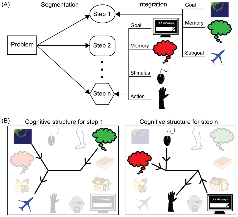
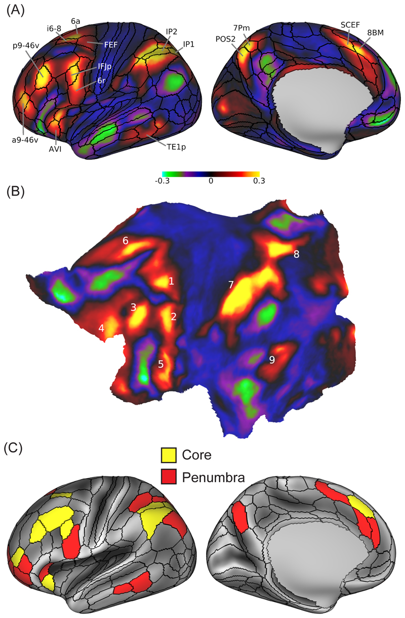
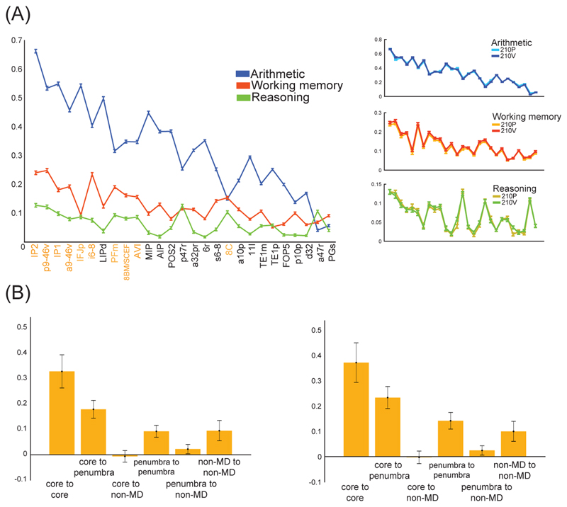
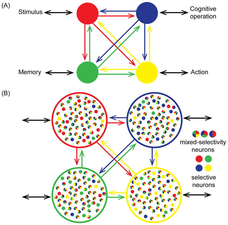
How does organized cognition arise from distributed brain activity? Recent analyses of fluid intelligence suggest a core process of cognitive focus and integration, organizing the components of a cognitive operation into the required computational structure. A cortical 'multiple-demand' (MD) system is closely linked to fluid intelligence, and recent imaging data define nine specific MD patches distributed across frontal, parietal, and occipitotemporal cortex. Wide cortical distribution, relative functional specialization, and strong connectivity suggest a basis for cognitive integration, matching electrophysiological evidence for binding of cognitive operations to their contents. Though still only in broad outline, these data suggest how distributed brain activity can build complex, organized cognition.
Keywords: attention; brain networks; cognitive control; intelligence; neural coding.
Copyright © 2020 The Authors. Published by Elsevier Ltd.. All rights reserved.
Figures
Similar articles
-
Graph lesion-deficit mapping of fluid intelligence.Brain. 2023 Jan 5;146(1):167-181. doi: 10.1093/brain/awac304. Brain. 2023. PMID: 36574957 Free PMC article.
-
Resting-State Network Patterns Underlying Cognitive Function in Bipolar Disorder: A Graph Theoretical Analysis.Brain Connect. 2020 Sep;10(7):355-367. doi: 10.1089/brain.2019.0709. Epub 2020 Jul 21. Brain Connect. 2020. PMID: 32458698 Free PMC article.
-
The structure of cognition: attentional episodes in mind and brain.Neuron. 2013 Oct 2;80(1):35-50. doi: 10.1016/j.neuron.2013.09.015. Epub 2013 Oct 2. Neuron. 2013. PMID: 24094101 Free PMC article. Review.
-
Intrinsic brain mapping of cognitive abilities: A multiple-dataset study on intelligence and its components.Neuroimage. 2025 Apr 1;309:121094. doi: 10.1016/j.neuroimage.2025.121094. Epub 2025 Feb 18. Neuroimage. 2025. PMID: 39978703
-
Brain connectivity and visual attention.Brain Connect. 2013;3(4):317-38. doi: 10.1089/brain.2012.0139. Epub 2013 Jun 8. Brain Connect. 2013. PMID: 23597177 Free PMC article. Review.
Cited by
-
Topographical functional correlates of interindividual differences in executive functions in young healthy twins.Brain Struct Funct. 2022 Jan;227(1):49-62. doi: 10.1007/s00429-021-02388-4. Epub 2021 Dec 4. Brain Struct Funct. 2022. PMID: 34865178 Free PMC article.
-
Robust Effects of Working Memory Demand during Naturalistic Language Comprehension in Language-Selective Cortex.J Neurosci. 2022 Sep 28;42(39):7412-7430. doi: 10.1523/JNEUROSCI.1894-21.2022. J Neurosci. 2022. PMID: 36002263 Free PMC article.
-
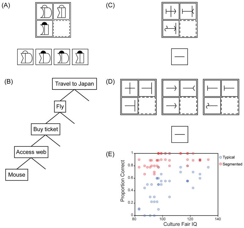
Cognitive segmentation and fluid reasoning in childhood.Q J Exp Psychol (Hove). 2023 Jun;76(6):1431-1444. doi: 10.1177/17470218221116054. Epub 2022 Aug 24. Q J Exp Psychol (Hove). 2023. PMID: 35848224 Free PMC article.
-
A middle ground where executive control meets semantics: the neural substrates of semantic control are topographically sandwiched between the multiple-demand and default-mode systems.Cereb Cortex. 2023 Apr 4;33(8):4512-4526. doi: 10.1093/cercor/bhac358. Cereb Cortex. 2023. PMID: 36130101 Free PMC article.
-
GABAergic inhibition in human hMT+ predicts visuo-spatial intelligence mediated through the frontal cortex.Elife. 2024 Oct 1;13:RP97545. doi: 10.7554/eLife.97545. Elife. 2024. PMID: 39352734 Free PMC article.
References
-
- Duncan J, et al. Intelligence and the frontal lobe: The organization of goal-directed behavior. Cognit Psychol. 1996;30:257–303. - PubMed
-
- Kane MJ, Engle RW. The role of prefrontal cortex in working memory capacity, executive attention, and general fluid intelligence: An individual-differences perspective. Psychonomic Bulletin and Review. 2002;9:637–671. - PubMed
-
- Unsworth N, Robison MK. A locus coeruleus-norepinephrine account of individual differences in working memory capacity and attention control. Psychonomic Bulletin and Review. 2017;24:1282–1311. - PubMed
Publication types
MeSH terms
Grants and funding
LinkOut - more resources
Full Text Sources
