. 2021;11(1):117-145.
doi: 10.1016/j.jcmgh.2020.07.011. Epub 2020 Aug 7.

Wnt-induced, TRP53-mediated Cell Cycle Arrest of Precursors Underlies Interstitial Cell of Cajal Depletion During Aging

Affiliations

Wnt-induced, TRP53-mediated Cell Cycle Arrest of Precursors Underlies Interstitial Cell of Cajal Depletion During Aging

Yujiro Hayashi et al. Cell Mol Gastroenterol Hepatol. 2021.

Abstract

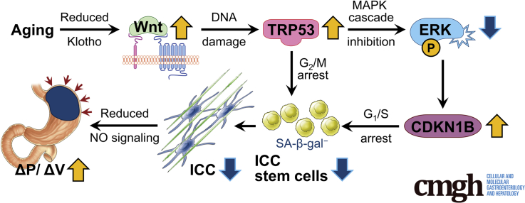
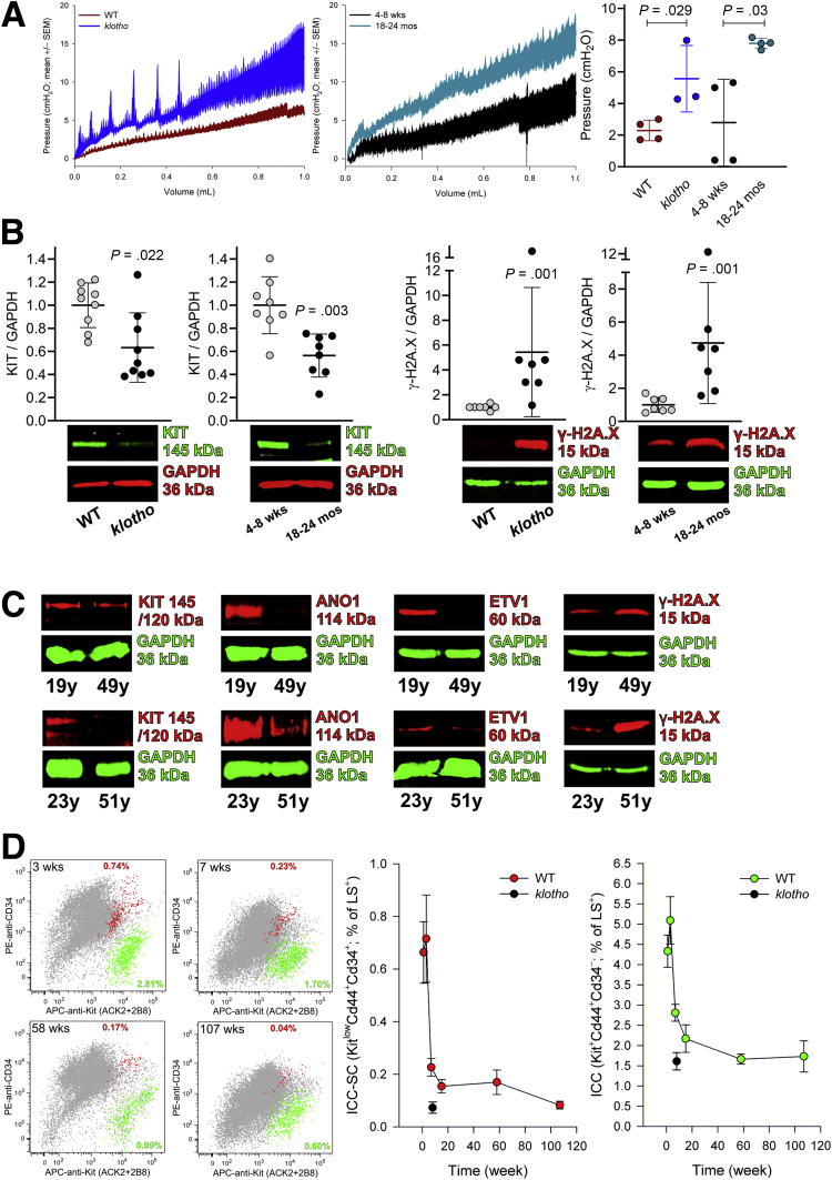
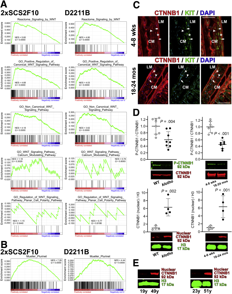
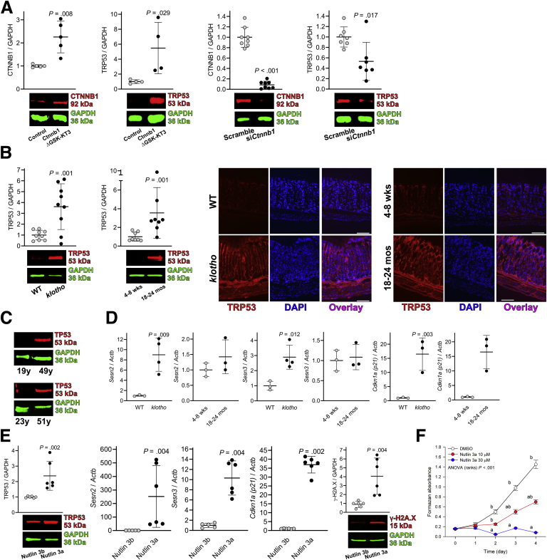
Background & aims: Gastric dysfunction in the elderly may cause reduced food intake, frailty, and increased mortality. The pacemaker and neuromodulator cells interstitial cells of Cajal (ICC) decline with age in humans, and their loss contributes to gastric dysfunction in progeric klotho mice hypomorphic for the anti-aging Klotho protein. The mechanisms of ICC depletion remain unclear. Klotho attenuates Wnt (wingless-type MMTV integration site) signaling. Here, we examined whether unopposed Wnt signaling could underlie aging-associated ICC loss by up-regulating transformation related protein TRP53 in ICC stem cells (ICC-SC).

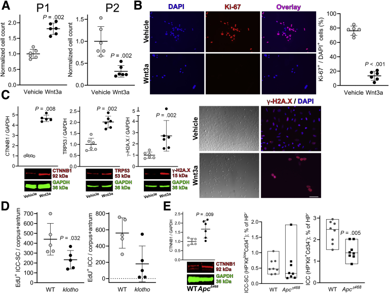
Methods: Mice aged 1-107 weeks, klotho mice, APCΔ468 mice with overactive Wnt signaling, mouse ICC-SC, and human gastric smooth muscles were studied by RNA sequencing, reverse transcription-polymerase chain reaction, immunoblots, immunofluorescence, histochemistry, flow cytometry, and methyltetrazolium, ethynyl/bromodeoxyuridine incorporation, and ex-vivo gastric compliance assays. Cells were manipulated pharmacologically and by gene overexpression and RNA interference.

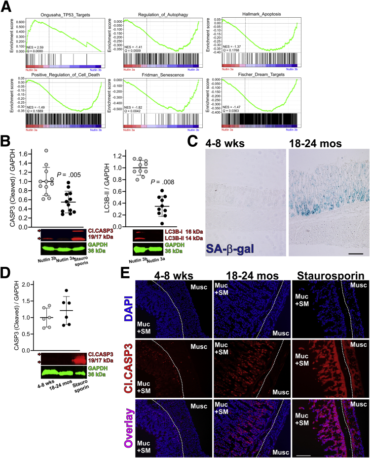
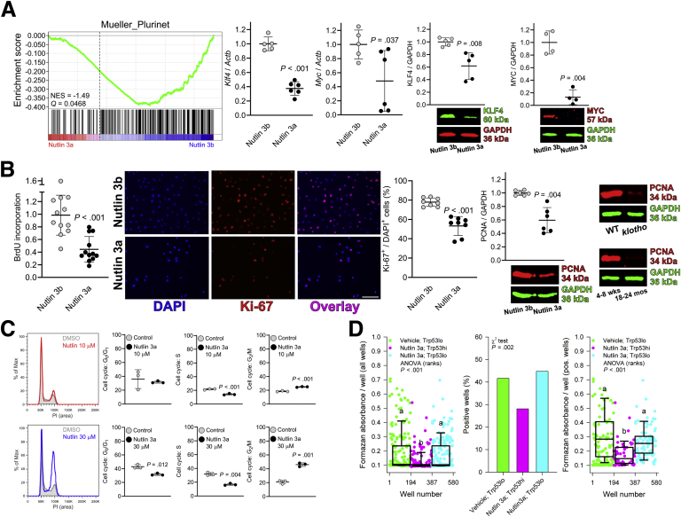
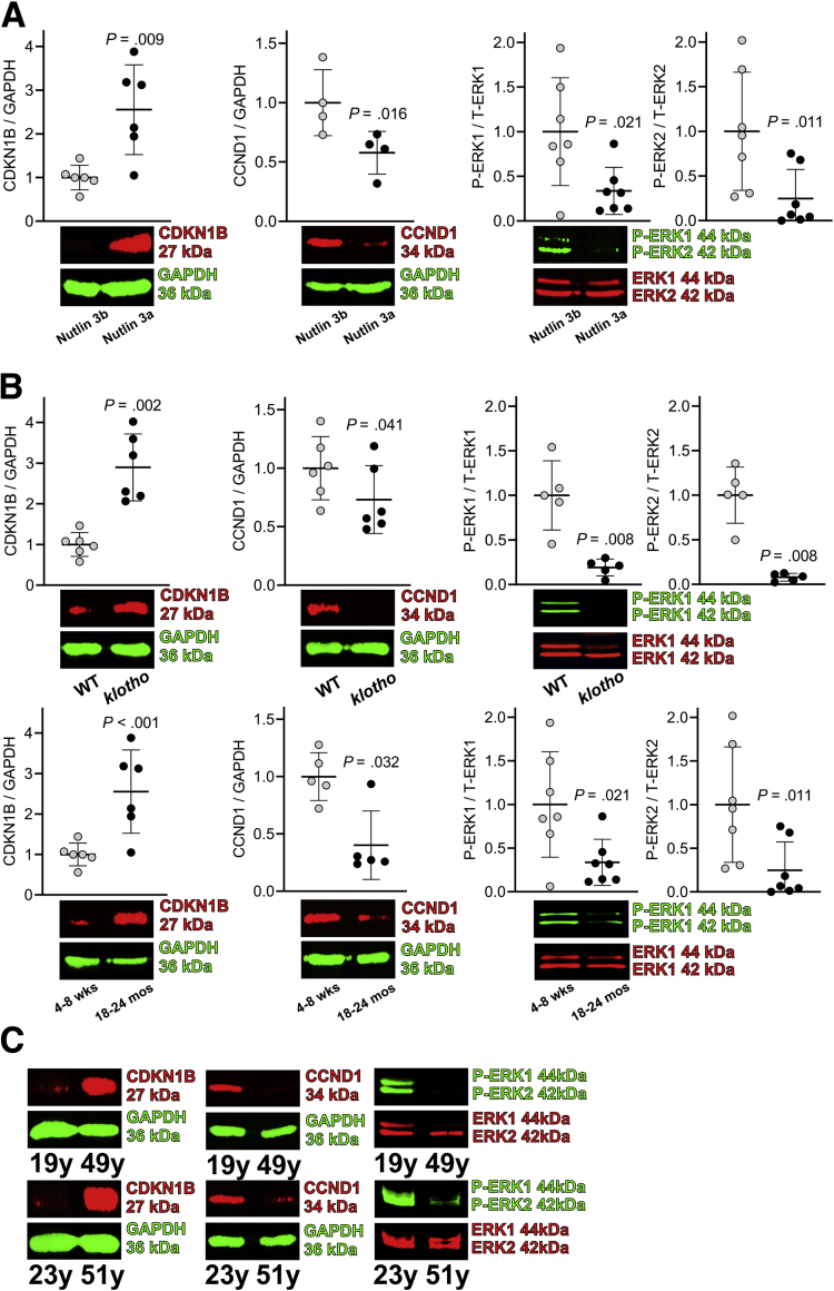
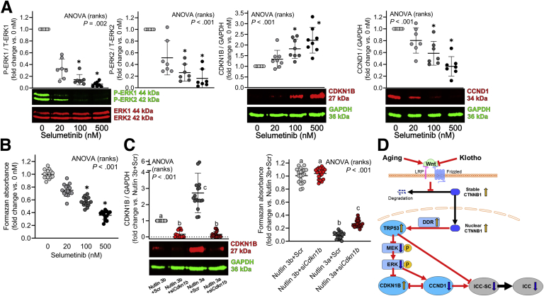
Results: The klotho and aged mice showed similar ICC loss and impaired gastric compliance. ICC-SC decline preceded ICC depletion. Canonical Wnt signaling and TRP53 increased in gastric muscles of klotho and aged mice and middle-aged humans. Overstimulated canonical Wnt signaling increased DNA damage response and TRP53 and reduced ICC-SC self-renewal and gastric ICC. TRP53 induction persistently inhibited G1/S and G2/M cell cycle phase transitions without activating apoptosis, autophagy, cellular quiescence, or canonical markers/mediators of senescence. G1/S block reflected increased cyclin-dependent kinase inhibitor 1B and reduced cyclin D1 from reduced extracellular signal-regulated kinase activity.

Conclusions: Increased Wnt signaling causes age-related ICC loss by up-regulating TRP53, which induces persistent ICC-SC cell cycle arrest without up-regulating canonical senescence markers.

Keywords: Compliance; Senescence; Stem Cell.


Figures

None
Graphical abstract
Figure 1
Figure 1
Age-related ICC and ICC-SC decline is associated with impaired gastric compliance. (A) Reduced gastric compliance of intact stomachs excised from 3 50- to 70-day-old klotho and 4 18- to 24-month-old C57BL/6 mice relative to age-matched WT (n = 4) and 4- to 8-week-old controls (n = 4), respectively (average traces). Stomachs were infused with 1 mL Krebs solution at 37°C at a rate of 0.1 mL/min while recording luminal pressure. P values are from Mann-Whitney rank sum tests. (B) Reduced KIT protein and increased γ-H2A.X (H2AXS139p) protein in the gastric tunica muscularis of klotho vs WT mice (n = 5–9) and old (18–24 months) vs young (4–8 weeks) mice (n = 7–9/group). GAPDH was used as a loading control. P values are from Mann-Whitney rank sum tests. (C) Down-regulated ICC-related proteins (KIT, ANO1, ETV1) and up-regulated γ-H2A.X in gastric corpus muscles of 49-year-old male patient and 51-year-old female patient vs 19-year-old male and 23-year-old female controls. (D) Gastric ICC (KIT+CD34 subpopulation; green) and ICC-SC (KITlowCD34+ subpopulation; red) frequencies assessed by flow cytometry in a mixture of male and female C57BL/6 (n = 31) and BALB/c (n = 20) mice between 1 and 107 weeks of age (6–14 mice/time point). Representative projections (left) and time course data (right) depicting age-associated reduction in ICC and ICC-SC frequencies are shown. Data points in black are ICC and ICC-SC frequencies in 7- to 10-week-old klotho mice shown for comparison.
Figure 2
Figure 2
The canonical Wnt signaling pathway is enriched in the ICC lineage and overactivated in the aging gastric tunica muscularis. (A) Enrichment of canonical and noncanonical Wnt pathways but not the calcium-modulating or planar cell polarity pathways in 2xSCS2F10 and D2211B ICC-SC lines analyzed by mRNA-seq and GSEA. Vertical lines indicate genes ranked by RPKM values. Gene sets with FDR Q value <0.25 and positive NES were considered significantly enriched. See further data and gene sets analyzed in Table 2 and Supplementary Table 1. (B) Enrichment of stemness-related genes in 2xSCS2F10 and D2211B ICC-SC lines by GSEA. See further data and gene sets analyzed in Table 3 and Supplementary Table 2. (C) Immunoreactivity for CTNNB1 (red), KIT (green), and DAPI (blue) in 5-μm cryosections of gastric tunica muscularis tissues from young and old mice (n = 3/group). Scale bars, 25 μm. CTNNB1 was expressed in ICC (arrows), KIT interstitial cells (arrowheads), and enteric neurons (asterisks) in both young and old mice. Note reduced KIT+ ICC in the old mouse. (D) CTNNB1 phosphorylation on Ser33/Ser37/Thr41 (P-CTNNB1) was reduced and nuclear CTNNB1 was increased in gastric corpus + antrum tissues of klotho mice vs WT controls and in old mice vs young controls (n = 4–8/group), indicating age-related overactivation of Wnt signaling. P values are from Mann-Whitney rank sum tests. (E) Up-regulated nuclear CTNNB1 protein in gastric corpus muscles of 49-year-old and 51-year-old patients vs 19-year-old and 23-year-old controls. CM, circular muscle; LM, longitudinal muscle.
Figure 3
Figure 3
Prolonged overactivation of canonical Wnt signaling causes paradoxical inhibition of ICC-SC proliferation. (A) 50,000 D2211B ICC-SC were cultured in the presence or absence of 30 ng/mL mouse recombinant Wnt3a (n = 12/group). Cell counts were determined when Wnt3a-treated cells reached confluence at 8 days of culturing (P1). Then 50,000 cells were re-plated (12 cultures/group) and counted when the controls reached confluence at 15 days (P2). ICC-SC counts in the Wnt3a-treated cultures were reduced after an initial increase. P values are from Mann-Whitney rank sum tests. (B) Reduced proliferation of D2211B ICC-SC detected by Ki-67 immunofluorescence after 15-day stimulation with Wnt3a (30 ng/mL). Nuclei were counterstained with DAPI. P value is from Mann-Whitney rank sum test. (C) Fifteen-day exposure of D2211B cells to 30 ng/mL Wnt3a up-regulated CTNNB1 and the DDR-associated proteins TRP53 and γ-H2A.X by WB and γ-H2A.X by immunofluorescence (n = 6/group). P values are from Mann-Whitney rank sum tests. (D) Reduced EdU+ proliferating ICC-SC and ICC detected in the gastric tunica muscularis of klotho vs WT mice by flow cytometry (n = 5/group). P values are from Mann-Whitney rank sum tests. (E) Up-regulated CTNNB1 and reduced ICC-SC and ICC in gastric tunica muscularis of APCΔ468 vs WT mice (n = 6–8/group). P values are from Mann-Whitney rank sum tests.
Figure 4
Figure 4
TRP53 is up-regulated in the aging gastric tunica muscularis and inhibits ICC-SC growth. (A) Overexpression of constitutively active Ctnnb1 in 2XSCS2F10 ICC-SC by nucleofection increased TRP53 protein levels (n = 5/group). The siRNA-mediated knockdown of Ctnnb1 down-regulated TRP53 protein expression in D2211B cells (n = 8/group). Efficacy of RNA interference against Ctnnb1 was verified by WB (n = 8/group). P values are from Mann-Whitney rank sum tests. (B) Left panels: increased TRP53 protein in klotho mouse gastric lysates compared with WT controls and in old vs young mice (n = 8–9/group). Right panels: immunoreactivity for TRP53 (red) and nuclear DAPI (blue) in cryosections of gastric tissues from klotho and WT control (n = 3/group) and young and old mice (n = 3/group). Note increased TRP53 protein in both the tunica mucosa and the tunica muscularis. P values are from Mann-Whitney rank sum tests. (C) Up-regulated TRP53 protein in the gastric corpus tunica muscularis of 49-year-old and 51-year-old patients vs 19-year-old and 23-year-old patients. (D) Increased expression of TRP53 transcriptional target genes Sesn2, Sesn3, and Cdkn1a (n = 3–4/group) by RT-qPCR in gastric tissues of klotho mice compared with WT controls. More modest changes were seen in old mice compared with young controls (n = 3–4/group). P values are from t tests. (E) Treatment with nutlin 3a, inhibitor of the TRP53-degrading E3 ubiquitin ligase MDM2 (30 μmol/L), of D2211B cells increased TRP53 and γ-H2A.X protein levels by WB (72 hours; n = 4/group) and the expression of TRP53 target genes Sesn2, Sesn3, and Cdkn1a by RT-qPCR (48 hours) vs treatment with the 150-fold less potent enantiomer nutlin 3b (n = 5–6/group). P values are from Mann-Whitney rank sum tests. (F) Nutlin 3a dose-dependently reduced D2211B proliferation by MTS assay (n = 7–8/group). P values are from Kruskal-Wallis one-way ANOVA (ANOVA on ranks). Groups not sharing the same superscript are different by multiple comparisons (P < .05; Tukey tests).
Figure 5
Figure 5
Cell death–, apoptosis-, autophagy-, senescence-, and quiescence-related pathways are not stimulated by TRP53 up-regulation in ICC-SC. (A) GSEA of total RNA-seq data showing enrichment of TRP53 target genes and depletion of cell death–, apoptosis-, autophagy-, senescence-, and quiescence (DREAM complex)-related genes in D2211B ICC-SC treated with nutlin 3a vs cells treated with nutlin 3b (30 μmol/L, 72 hours; n = 3/group). Vertical lines indicate genes. Negative NES and FDA Q <0.25 indicate significant depletion. See all data and gene sets analyzed in Tables 4 and 5 and in Supplementary Tables 3 and 4. (B) Nutlin 3a (30 μmol/L, 72 hours) in D2211B cells reduced CL.CASP3 (n = 12/group) and light chain 3B isoform II (LC3B-II) levels vs nutlin 3b (n = 7/group). Staurosporin (3 μmol/L, 24 hours) was used as a positive control for apoptosis induction. P values are from Mann-Whitney rank sum tests. (C) Senescent cells detected by SA-β-Gal activity were increased in the gastric mucosa of old (18–24 months) vs young mice (4–8 weeks). No SA-β-gal activity was evident in the gastric muscle layers of either old or young mice. (D and E) Apoptosis detected in the gastric corpus + antrum tunica muscularis by CL.CASP3 immunoblotting (D) and in the gastric corpus + antrum (full thickness) by immunohistochemistry (E) in old (18–24 months) vs young mice (4–8 weeks). CL.CASP3 activity was low in the gastric muscle layers of both old and young mice and did not increase with age. Positive controls were exposed to staurosporin (3 μmol/L, 24 hours; n = 6/group). Data in D were analyzed by Mann-Whitney rank sum test.
Figure 6
Figure 6
TRP53 activation inhibits ICC-SC growth by reducing cell proliferation and clonogenicity via down-regulation of self-renewal genes and cell cycle arrest. (A) Left panel: GSEA of total RNA-seq data showing down-regulation of stemness-related genes in D2211B ICC-SC treated with nutlin 3a vs cells treated with nutlin 3b (30 μmol/L, 72 hours; n = 3/group). Vertical lines indicate genes ranked by signal-to-noise ratios. Negative NES and FDR Q <0.25 indicate significant depletion in nutlin 3a–treated group. See all data and gene sets analyzed in Table 4 and Supplementary Table 2. Right panels: reduced mRNA and protein levels of stemness genes KLF4 and MYC. P values are from Mann-Whitney rank sum tests. (B) Nutlin 3a (30 μmol/L, 72 hours) reduced D2211B proliferation by BrdU incorporation (n = 12/group), Ki-67 immunofluorescence (n = 10/group), and PCNA protein expression (n = 8/group). PCNA expression was also reduced in klotho and old mice vs their respective controls (n = 6/group). P values are from Mann-Whitney rank sum tests. (C) Cell cycle arrest detected by PI flow cytometric assay in D2211B cells treated with 30 μmol/L or 10 μmol/L nutlin 3a for 72 hours (n = 3/group). P values are from t tests applied to arcsine square root transformed data. (D) Nutlin 3a–treated (10 μmol/L, 3 days), clonally sorted, TRP53high 2xSCS2F10 ICC-SC lentivirally transduced with the pGreenFire-p53-mCMV-EF1α-Puro Transcriptional Reporter displayed reduced clonogenicity and proliferation by MTS assay than vehicle-treated or nutlin 3a–treated, TRP53low cells (192 wells/group). In the left and right panels, P values are from Kruskal-Wallis one-way ANOVA (ANOVA on ranks). Groups not sharing the same superscript are different by multiple comparisons (left panel, P < .05, Tukey tests; right panel, P < .05, Dunn’s method). Proportions in the middle panel were analyzed by χ2 test. PCNA, proliferating cell nuclear antigen.
Figure 7
Figure 7
Increased CDKN1B, reduced CCND1, and reduced ERK activation in nutlin 3a–treated ICC-SC and during aging. (A) Nutlin 3a treatment (30 μmol/L, 72 hours) in D2211B cells up-regulated CDKN1B protein levels, down-regulated CCND1 protein levels, and reduced ERK1/2 phosphorylation by WB (n = 4–7/group). P values are from Mann-Whitney rank sum tests. (B) Increased CDKN1B protein, reduced CCND1 protein, and reduced ERK1/2 phosphorylation in klotho mouse gastric lysates compared with WT controls and in old vs young mice (n = 5–8/group). P values are from Mann-Whitney rank sum tests. (C) Up-regulated CDKN1B protein, reduced CCDN1 protein, and reduced ERK1/2 phosphorylation in the gastric corpus muscles of 49-year-old and 51-year-old patients vs 19-year-old and 23-year-old controls.
Figure 8
Figure 8
TRP53 inhibits ICC-SC proliferation via the ERK-CDKN1B-CCND1 pathway. (A) The mitogen-activated protein kinase kinase 1/2 (MEK1/2) inhibitor selumetinib applied for 3 days dose-dependently reduced ERK phosphorylation, increased CDKN1B protein levels, and reduced CCND1 protein by WB (n = 7–8/group) in D2211B ICC-SC. P values are from Kruskal-Wallis one-way ANOVA (ANOVA on ranks). ∗P < .05 vs vehicle control by post hoc multiple comparisons (Dunn’s method). (B) Selumetinib also dose-dependently inhibited ICC-SC viability by MTS assay (n = 15/group). P value is from Kruskal-Wallis one-way ANOVA (ANOVA on ranks). ∗P < .05 vs vehicle control by post hoc multiple comparisons (Dunn’s method). (C) siRNA-mediated knockdown of Cdkn1b, verified by WB, mitigated the nutlin 3a–induced reduction in D2211B growth by MTS assay (n = 20/group). P values are from Kruskal-Wallis one-way ANOVA (ANOVA on ranks). Groups not sharing the same superscript are different by multiple comparisons (P < .05, Tukey tests). (D) Proposed mechanisms of ICC-SC depletion leading to ICC loss during aging. Aging causes overactive Wnt signaling in part by reducing Klotho protein levels. Increased Wnt signaling increases TRP53 protein levels by stabilizing CTNNB1 and promoting its nuclear localization and by inducing DDR. TRP53 causes ICC depletion in part by inhibiting ICC-SC proliferation via G2/M arrest. TRP53 also inhibits ERK1/2 phosphorylation (P), decreasing CCND1 and increasing CDKN1B protein levels. Reduced CCND1, likely together with other G1/S cyclins, down-regulates ICC-SC proliferation and self-renewal by interfering with S phase entry.

Similar articles

Cited by

References

    1. Kennedy B.K., Berger S.L., Brunet A., Campisi J., Cuervo A.M., Epel E.S., Franceschi C., Lithgow G.J., Morimoto R.I., Pessin J.E., Rando T.A., Richardson A., Schadt E.E., Wyss-Coray T., Sierra F. Geroscience: linking aging to chronic disease. Cell. 2014;159:709–713. - PMC - PubMed
    1. Camilleri M., Cowen T., Koch T.R. Enteric neurodegeneration in ageing. Neurogastroenterol Motil. 2008;20:185–196. - PubMed
    1. Bhutto A., Morley J.E. The clinical significance of gastrointestinal changes with aging. Curr Opin Clin Nutr Metab Care. 2008;11:651–660. - PubMed
    1. Salles N. Is stomach spontaneously ageing? pathophysiology of the ageing stomach. Best Pract Res Clin Gastroenterol. 2009;23:805–819. - PubMed
    1. Parker B.A., Chapman I.M. Food intake and ageing: the role of the gut. Mech Ageing Dev. 2004;125:859–866. - PubMed

Publication types

Substances