Review
. 2021 Mar:81:100886.
doi: 10.1016/j.preteyeres.2020.100886. Epub 2020 Aug 6.

Role of glia in optic nerve

Affiliations
Review

Role of glia in optic nerve

Meysam Yazdankhah et al. Prog Retin Eye Res. 2021 Mar.

Abstract

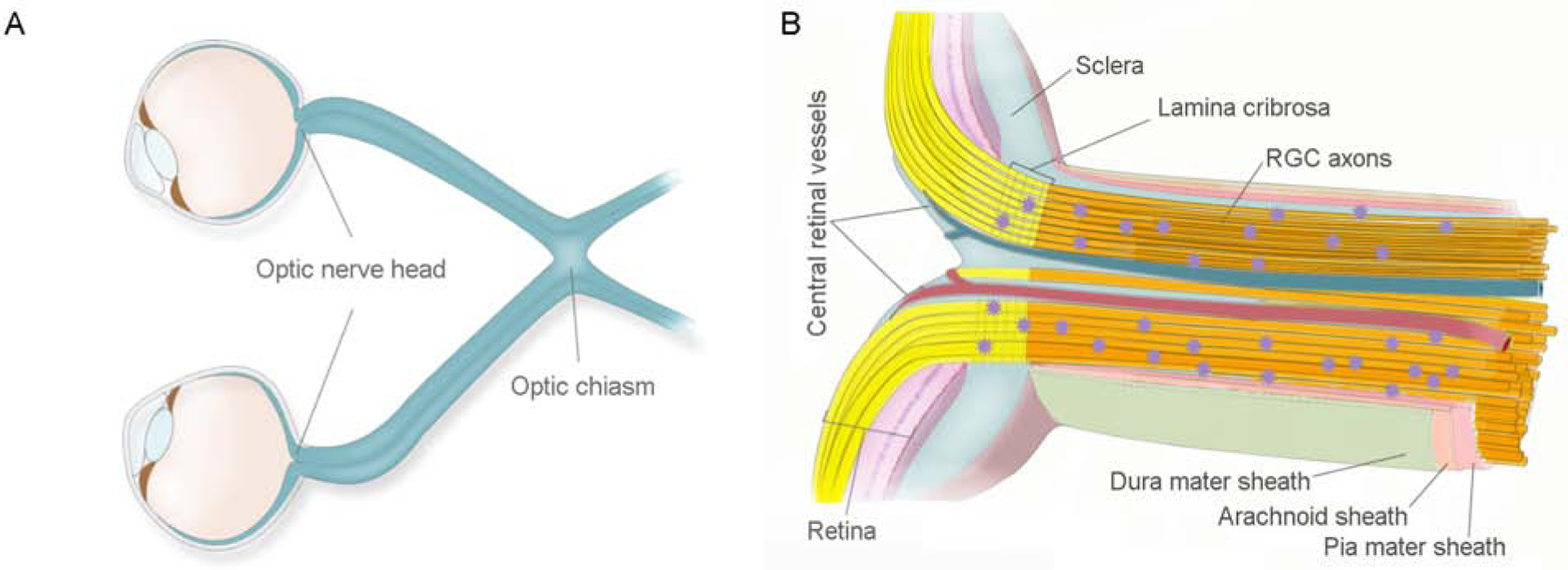
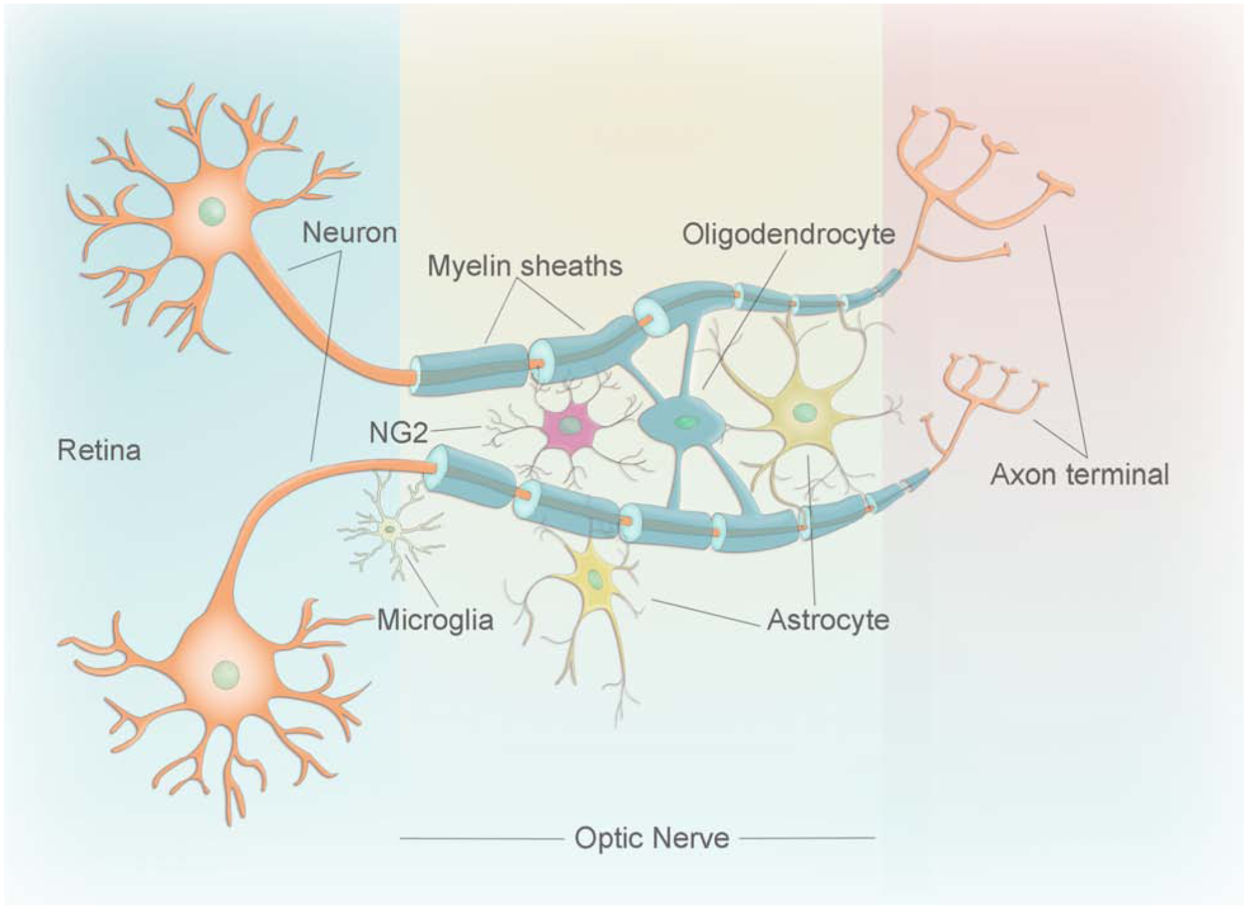
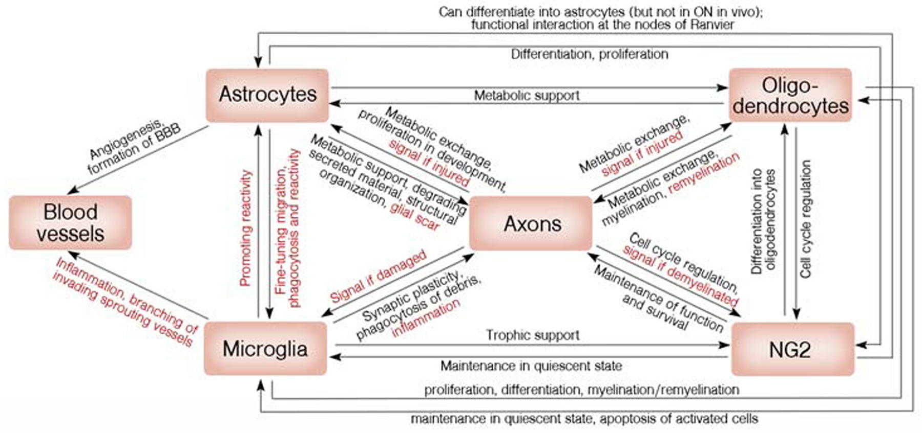
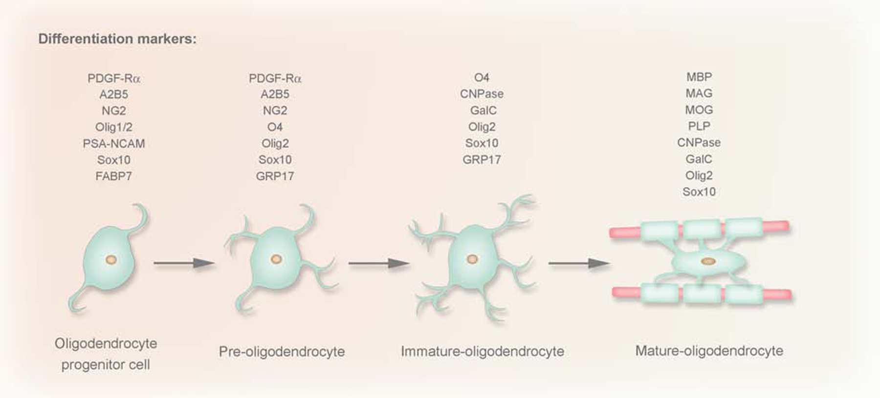
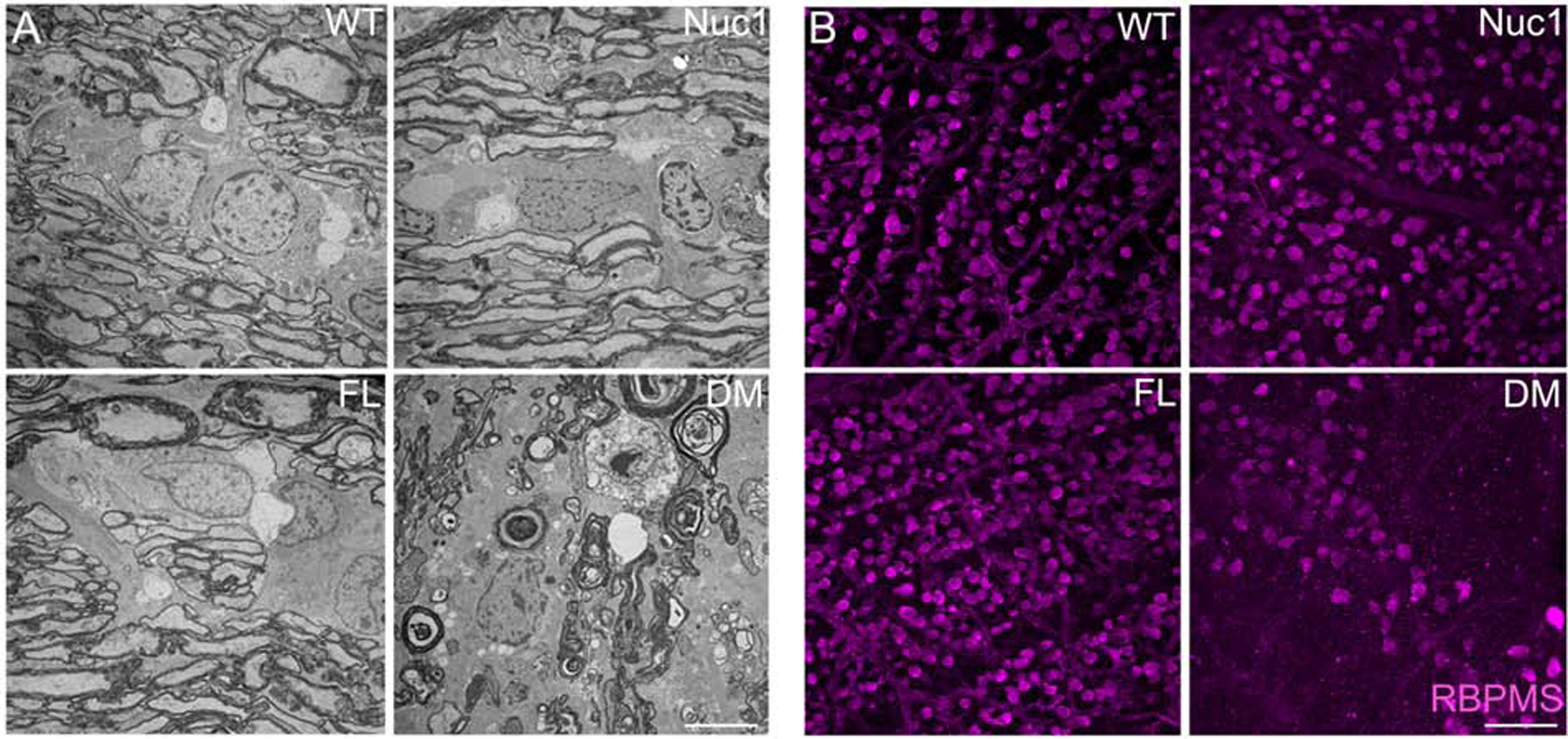
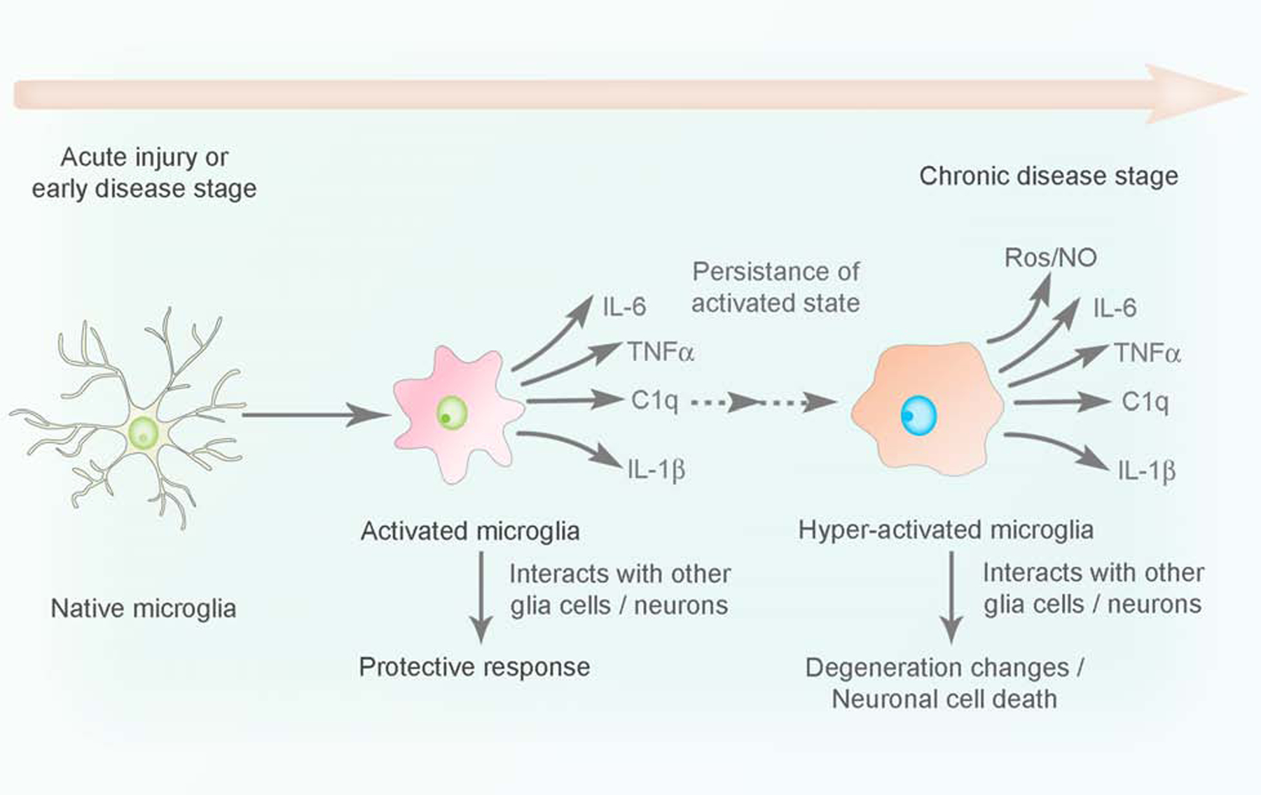
Glial cells are critically important for maintenance of neuronal activity in the central nervous system (CNS), including the optic nerve (ON). However, the ON has several unique characteristics, such as an extremely high myelination level of retinal ganglion cell (RGC) axons throughout the length of the nerve (with virtually all fibers myelinated by 7 months of age in humans), lack of synapses and very narrow geometry. Moreover, the optic nerve head (ONH) - a region where the RGC axons exit the eye - represents an interesting area that is morphologically distinct in different species. In many cases of multiple sclerosis (demyelinating disease of the CNS) vision problems are the first manifestation of the disease, suggesting that RGCs and/or glia in the ON are more sensitive to pathological conditions than cells in other parts of the CNS. Here, we summarize current knowledge on glial organization and function in the ON, focusing on glial support of RGCs. We cover both well-established concepts on the important role of glial cells in ON health and new findings, including novel insights into mechanisms of remyelination, microglia/NG2 cell-cell interaction, astrocyte reactivity and the regulation of reactive astrogliosis by mitochondrial fragmentation in microglia.

Keywords: Astrocytes; Glia; Microglia; Neuron glial 2 (NG2) cells; Oligodendrocytes; Optic nerve (ON).


Figures

Figure 1:
Figure 1:. The structure of the optic nerve A.
Optic nerve is a white matter tract of the brain that connects the eye (from the optic nerve head end) with the brain. The left and right optic nerves intersect at the chiasm, which is an X-shaped structure that is located in the forebrain, in front of the hypothalamus. B. The optic nerve is composed of myelinated axons of the retinal ganglion cells (RGCs) (yellow bundles) and interstitial glial cells (purple stars). Numerous septa of neuroglia, plus elastic and collagenous fibers divide the nerve into sharply defined bundles that are encapsulated by the three meningeal layers of the brain (dura, arachnoid, and pia mater). The main structural element of the optic nerve head is the dense collagenous lamina cribrosa that acts as a pressure barrier between the intraocular and retrobulbar spaces.
Figure 2:
Figure 2:. Schematic diagram of glial cells in the optic nerve.
Cartoon showing the different types of glial cells (astrocytes, oligodendrocytes, NG2 cells, and microglia) in the optic nerve and their association with the neuron.
Figure 3.
Figure 3.. Schematic representation of the simplified interactions between glial cells in the optic nerve.
Glial cells work as a team to ensure both healthy homeostasis (black annotations) and response to insults (red) in the optic nerve.
Figure 4:
Figure 4:. Four different phases of oligodendrocyte (OL) maturation in the oligodendroglial lineage: OPC, pre-OLs, immature OLs and mature OLs.
These phases were established based on the increasingly complex morphology and the expression pattern of well-defined markers. CNPase, 2′,3′-cyclic nucleotide 3′-phosphodiesterase; FABP, fatty-acid-binding proteins; GalC, galactocerebroside C; MAG, myelin associated glycoprotein; MBP, myelin basic protein; MOG, myelin OL glycoprotein;Olig1/2 two basic helix-loop-helix transcription factors; PDGF-Rα, platelet-derived growth factor; PLP, proteolipid protein; PSA-NCAM, polysialic acid-neural cell adhesion molecule.
Figure 5:
Figure 5:. Rats homozygous for both Cryba1 and Bckdk mutations exhibit very serious optic nerve degeneration with profound demyelination.
To understand the roles of Bckdk and Cryba1 in optic nerve survival, the optic nerves and retinas were dissected from wild type (WT), Nuc1 (mutation in Cryba1), FL (mutation in Bckdk, named frogleg (FL) because of a severe splaying of the hind limbs) and DM (double mutant, mutations in both Cryba1 and Bckdk) rats. (A) TEM revealed a severe optic nerve degeneration in the DM rats compared to other genotypes at 4 months of age. Bar: 5 μm. (B) Immunohistochemistry of retinal flat mounts against RBPMS (Anti-RNA binding protein with multiple splicing) showed degeneration of RGCs in 4 month old DM rats relative to other genotypes. Bar: 100 μm.
Figure 6:
Figure 6:. Microglia activation during disease progression.
Naïve microglia undergo inflammatory transition to an activated state (releasing inflammatory cytokines, chemokines) to confer protection during acute infection or tissue injury in the CNS. However, persistent activation of microglia upon stress stimuli rendered by genetic predisposition or environmental factors, leads to a state of hyper activation associated with an uncontrolled release of pro-inflammatory mediators (chronic inflammation), which in turn causes neuronal damage in the CNS.

References

    1. Agapova OA, Kaufman PL, Lucarelli MJ, Gabelt BAT, Hernandez MR, 2003. Differential expression of matrix metalloproteinases in monkey eyes with experimental glaucoma or optic nerve transection. Brain Res, 967, 132–143. - PubMed
    1. Agapova OA, Ricard CS, Salvador-Silva M, Hernandez MR, 2001. Expression of matrix metalloproteinases and tissue inhibitors of metalloproteinases in human optic nerve head astrocytes. Glia. 33, 205–216. - PubMed
    1. Al-Gayyar MM, Elsherbiny NM, 2013. Contribution of TNF-α to the development of retinal neurodegenerative disorders. Eur. Cytokine. Netw 24, 27–36. - PubMed
    1. Alkozi HA, Navarro G, Franco R, Pintor J, 2019. Melatonin and the control of intraocular pressure. Prog Retin Eye Res. 25, 100798. - PubMed
    1. Allen A, Messier C, 2013. Plastic changes in the astrocyte GLUT1 glucose transporter and beta-tubulin microtubule protein following voluntary exercise in mice. Behav Brain Res 240: 95–102. - PubMed

Publication types