

Sexually Dimorphic Crosstalk at the Maternal-Fetal Interface
- PMID: 32772088
- PMCID: PMC7571453
- DOI: 10.1210/clinem/dgaa503
Sexually Dimorphic Crosstalk at the Maternal-Fetal Interface
Abstract
Context: Crosstalk through receptor ligand interactions at the maternal-fetal interface is impacted by fetal sex. This affects placentation in the first trimester and differences in outcomes. Sexually dimorphic signaling at early stages of placentation are not defined.
Objective: Investigate the impact of fetal sex on maternal-fetal crosstalk.
Design: Receptors/ligands at the maternal-fetal surface were identified from sexually dimorphic genes between fetal sexes in the first trimester placenta and defined in each cell type using single-cell RNA-Sequencing (scRNA-Seq).
Setting: Academic institution.
Samples: Late first trimester (~10-13 weeks) placenta (fetal) and decidua (maternal) from uncomplicated ongoing pregnancies.
Main outcome measures: Transcriptomic profiling at tissue and single-cell level; immunohistochemistry of select proteins.
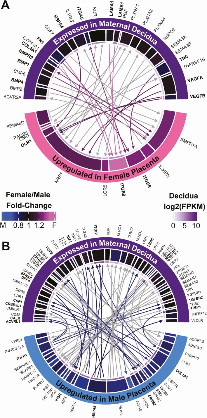
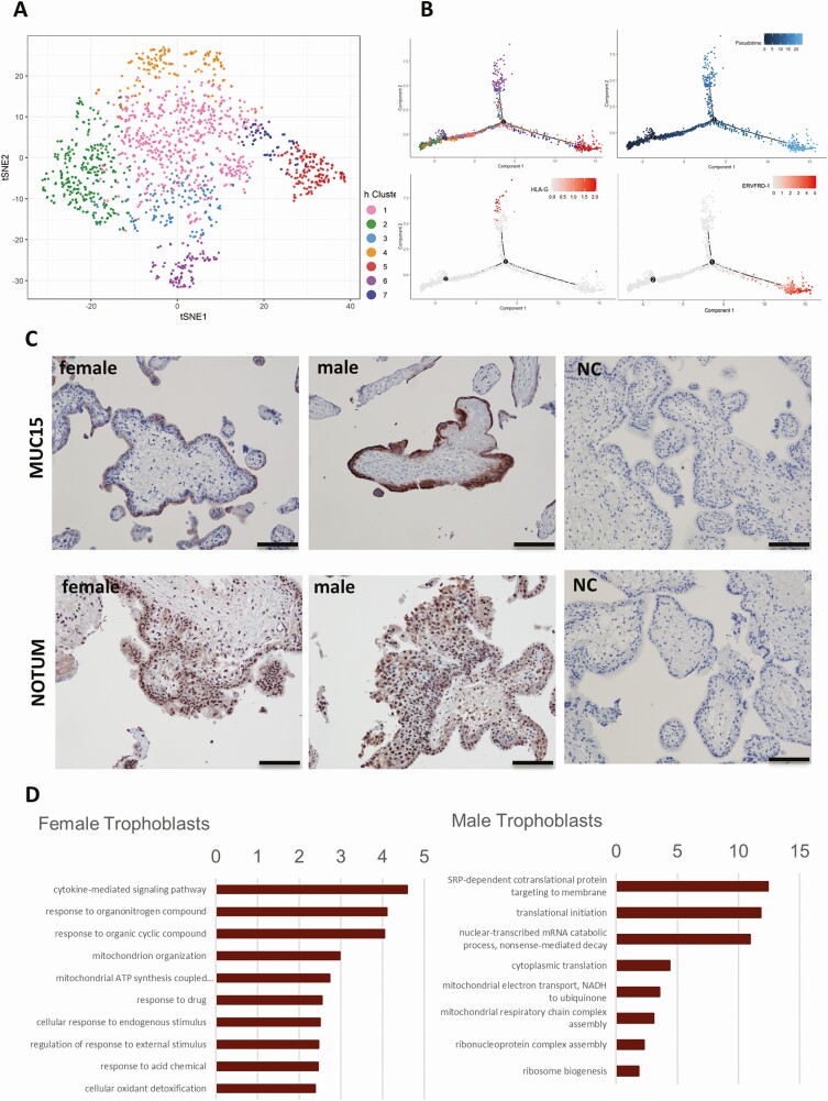
Results: We identified 91 sexually dimorphic receptor-ligand pairs across the maternal-fetal interface. We examined fetal sex differences in 5 major cell types (trophoblasts, stromal cells, Hofbauer cells, antigen-presenting cells, and endothelial cells). Ligands from the CC family chemokine ligand (CCL) family were most highly representative in females, with their receptors present on the maternal surface. Sexually dimorphic trophoblast transcripts, Mucin-15 (MUC15) and notum, palmitoleoyl-protein carboxylesterase (NOTUM) were also most highly expressed in syncytiotrophoblasts and extra-villous trophoblasts respectively. Gene Ontology (GO) analysis using sexually dimorphic genes in individual cell types identified cytokine mediated signaling pathways to be most representative in female trophoblasts. Upstream analysis demonstrated TGFB1 and estradiol to affect all cell types, but dihydrotestosterone, produced by the male fetus, was an upstream regulator most significant for the trophoblast population.
Conclusions: Maternal-fetal crosstalk exhibits sexual dimorphism during placentation early in gestation.
Keywords: first trimester placenta; human pregnancy; placenta cell types; receptor-ligand; sex differences; single-cell RNA sequencing.
© The Author(s) 2020. Published by Oxford University Press on behalf of the Endocrine Society. All rights reserved. For permissions, please e-mail: journals.permissions@oup.com.
Figures

Comment in
-
Let's Talk About Sex: Placentas' Central Role in Sexually Dimorphic Responses to the Maternal Milieu.J Clin Endocrinol Metab. 2020 Dec 1;105(12):e4973-4. doi: 10.1210/clinem/dgaa683. J Clin Endocrinol Metab. 2020. PMID: 32966581 Free PMC article. No abstract available.
Similar articles
-
Revealing the molecular landscape of human placenta: a systematic review and meta-analysis of single-cell RNA sequencing studies.Hum Reprod Update. 2024 Jul 1;30(4):410-441. doi: 10.1093/humupd/dmae006. Hum Reprod Update. 2024. PMID: 38478759 Free PMC article.
-
Sexually dimorphic DNA methylation and gene expression patterns in human first trimester placenta.Biol Sex Differ. 2024 Aug 16;15(1):63. doi: 10.1186/s13293-024-00629-9. Biol Sex Differ. 2024. PMID: 39152463 Free PMC article.
-
High-throughput mRNA sequencing of human placenta shows sex differences across gestation.Placenta. 2024 May;150:8-21. doi: 10.1016/j.placenta.2024.03.005. Epub 2024 Mar 21. Placenta. 2024. PMID: 38537412 Free PMC article.
-
HLA Class I protein expression in the human placenta.Early Pregnancy (Cherry Hill). 2001 Jan;5(1):67-9. Early Pregnancy (Cherry Hill). 2001. PMID: 11753519
-
Current approaches and developments in transcript profiling of the human placenta.Hum Reprod Update. 2020 Nov 1;26(6):799-840. doi: 10.1093/humupd/dmaa028. Hum Reprod Update. 2020. PMID: 33043357 Free PMC article. Review.
Cited by
-
Revealing the molecular landscape of human placenta: a systematic review and meta-analysis of single-cell RNA sequencing studies.Hum Reprod Update. 2024 Jul 1;30(4):410-441. doi: 10.1093/humupd/dmae006. Hum Reprod Update. 2024. PMID: 38478759 Free PMC article.
-
Sexually dimorphic DNA methylation and gene expression patterns in human first trimester placenta.Biol Sex Differ. 2024 Aug 16;15(1):63. doi: 10.1186/s13293-024-00629-9. Biol Sex Differ. 2024. PMID: 39152463 Free PMC article.
-
Sex-specific phenotypical, functional and metabolic profiles of human term placenta macrophages.Biol Sex Differ. 2024 Oct 17;15(1):80. doi: 10.1186/s13293-024-00652-w. Biol Sex Differ. 2024. PMID: 39420346 Free PMC article.
-
Leveraging chorionic villus biopsies for the derivation of patient-specific trophoblast stem cells.Commun Biol. 2025 Jul 1;8(1):964. doi: 10.1038/s42003-025-08393-1. Commun Biol. 2025. PMID: 40596474 Free PMC article.
-
Single cell profiling at the maternal-fetal interface reveals a deficiency of PD-L1+ non-immune cells in human spontaneous preterm labor.Sci Rep. 2023 May 16;13(1):7903. doi: 10.1038/s41598-023-35051-5. Sci Rep. 2023. PMID: 37193763 Free PMC article.
References
-
- Basso O, Olsen J. Sex ratio and twinning in women with hyperemesis or pre-eclampsia. Epidemiology. 2001;12(6):747-749. - PubMed
-
- Melamed N, Yogev Y, Glezerman M. Fetal gender and pregnancy outcome. J Matern Fetal Neonatal Med. 2010;23(4):338-344. - PubMed
-
- Ricart W, López J, Mozas J, et al. ; Spanish Group for the study of the impact of Carpenter and Coustan GDM thresholds . Maternal glucose tolerance status influences the risk of macrosomia in male but not in female fetuses. J Epidemiol Community Health. 2009;63(1):64-68. - PubMed
-
- Astolfi P, Zonta LA. Risks of preterm delivery and association with maternal age, birth order, and fetal gender. Hum Reprod. 1999;14(11):2891-2894. - PubMed
-
- Zeitlin J, Saurel-Cubizolles MJ, De Mouzon J, et al. Fetal sex and preterm birth: are males at greater risk? Hum Reprod. 2002;17(10):2762-2768. - PubMed
Publication types
MeSH terms
Grants and funding
LinkOut - more resources
Full Text Sources
Molecular Biology Databases
Miscellaneous
