. 2021 Jan;63(1):91-98.
doi: 10.1007/s00234-020-02508-7. Epub 2020 Aug 8.

Automated 3D MRI rendering of the craniofacial skeleton: using ZTE to drive the segmentation of black bone and FIESTA-C images

Affiliations

Automated 3D MRI rendering of the craniofacial skeleton: using ZTE to drive the segmentation of black bone and FIESTA-C images

Karen A Eley et al. Neuroradiology. 2021 Jan.

Abstract

Purpose: Automated bone segmentation from MRI datasets would have a profound impact on clinical utility, particularly in the craniofacial skeleton where complex anatomy is coupled with radiosensitive organs. Techniques such as gradient echo black bone (GRE-BB) and short echo time (UTE, ZTE) have shown potential in this quest. The objectives of this study were to ascertain (1) whether the high-contrast of zero echo time (ZTE) could drive segmentation of high-resolution GRE-BB data to enhance 3D-output and (2) if these techniques could be extrapolated to ZTE driven segmentation of a routinely used non bone-specific sequence (FIESTA-C).

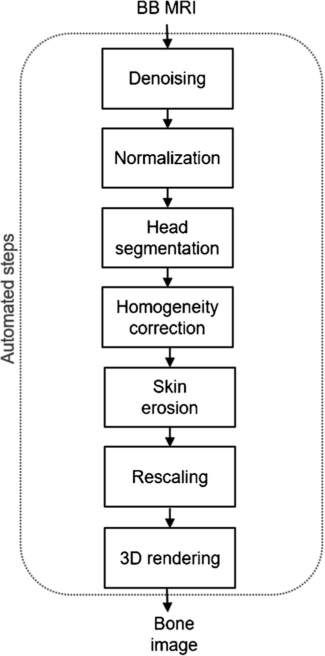
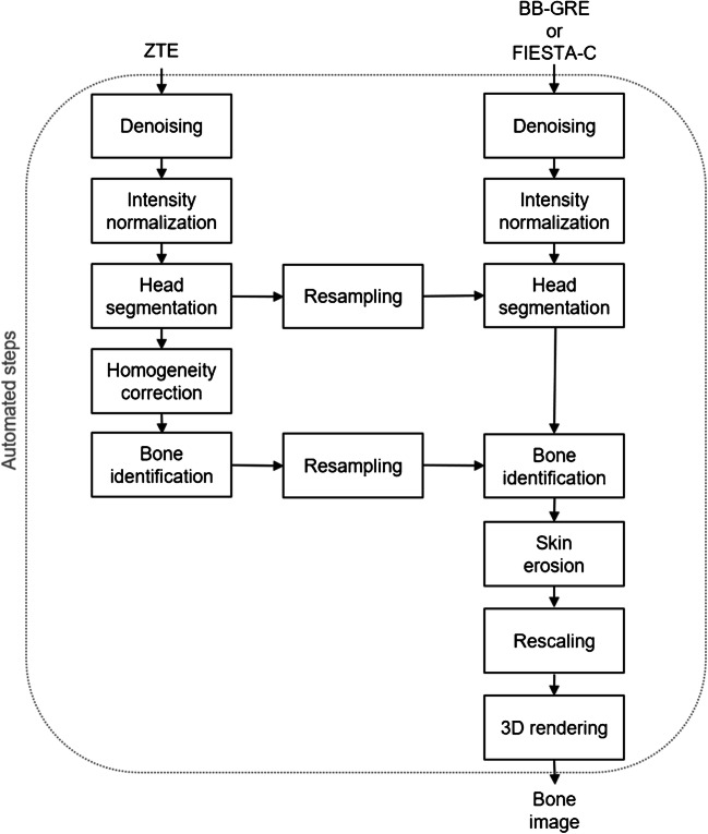
Methods: Eleven adult volunteers underwent 3T MRI examination with sequential acquisition of ZTE, GRE-BB and FIESTA-C imaging. Craniofacial bone segmentation was performed using a fully automated segmentation algorithm. Segmentation was completed individually for GRE-BB and a modified version of the algorithm was subsequently implemented, wherein the bone mask yielded by ZTE segmentation was used to initialise segmentation of GRE-BB. The techniques were subsequently applied to FIESTA-C datasets. The resulting 3D reconstructions were evaluated for areas of unexpected bony defects and discrepancies.

Results: The automated segmentation algorithm yielded acceptable 3D outputs for all GRE-BB datasets. These were enhanced with the modified algorithm using ZTE as a driver, with improvements in areas of air/bone interface and dense muscular attachments. Comparable results were obtained with ZTE+FIESTA-C.

Conclusion: Automated 3D segmentation of the craniofacial skeleton is enhanced through the incorporation of a modified segmentation algorithm utilising ZTE. These techniques are transferrable to FIESTA-C imaging which offers reduced acquisition time and therefore improved clinical utility.

Keywords: Facial bones; Image processing, computer-assisted]; Magnetic resonance imaging; Skull; Three-dimensional imaging.


Conflict of interest statement

Dr. Delso is currently employed by GE Healthcare.

Figures

Fig. 1
Fig. 1
Block diagram of the automated bone rendering algorithm, relying only on black bone images (BB-GRE)
Fig. 2
Fig. 2
Block diagram of the new bone rendering algorithm, relying on low-resolution ZTE data to enhance the depiction of high-resolution data (BB-GRE or FIESTA-C)
Fig. 3
Fig. 3
Axial acquisition of BB, ZTE and FIESTA-C demonstrating the inherent low-resolution imaging of ZTE when compared with BB and FIESTA-C
Fig. 4
Fig. 4
Automated 3D reconstruction of the craniofacial skeleton in adult volunteers to highlight differences between BB and BB+ZTE segmentation algorithms. Note the improved accuracy of BB+ZTE in terms of removal of cartilaginous and muscular structures. Areas of discrepancy are noted overlying the frontal sinus (a), and lateral orbital wall (b) and skull vertex (c) on BB images, with some improvement seen on the modified BB+ZTE results
Fig. 5
Fig. 5
Post-processed raw BB coronal imaging and 3D volume rendering of the dataset shown in Fig. 4C. This demonstrates the absence of bony defects at the skull vertex, highlighting that these defects are most likely a post-processing phenomenon and thus resolvable with future refinement of the technique
Fig. 6
Fig. 6
Fine manipulation of the final 3D-rendered imaging is possible by the end-user utilising software such as Osirix. Shown here are a range of 3D imaging from the same processed BB-ZTE dataset
Fig. 7
Fig. 7
Automated 3D reconstruction of the craniofacial skeleton in adult volunteers using FIESTA-C and a modified algorithm with ZTE+FIESTA-C

References

    1. Kramer M, Herzau B, Reichenbach JR. Segmentation and visualization of human cranial bone by T2* approximation using ultra-short echo time (UTE) magnetic resonance imaging. Z Med Phys. 2020;30:51–59. doi: 10.1016/j.zemedi.2019.06.003. - DOI - PubMed
    1. Eley KA, McIntyre AG, Watt-Smith SR, Golding SJ. “Black bone” MRI: a partial flip angle technique for radiation reduction in craniofacial imaging. Br J Radiol. 2012;85(1011):272–278. doi: 10.1259/bjr/95110289. - DOI - PMC - PubMed
    1. Eley KA, Watt-Smith SR, Sheerin F, Golding SJ. “Black bone” MRI: a potential alternative to CT with three-dimensional reconstruction of the craniofacial skeleton in the diagnosis of craniosynostosis. Eur Radiol. 2014;24(10):2417–2426. doi: 10.1007/s00330-014-3286-7. - DOI - PubMed
    1. Eley KA, Watt-Smith SR, Golding SJ. Three-dimensional reconstruction of the craniofacial skeleton with gradient echo magnetic resonance imaging (“black bone”): what is currently possible? J Craniofac Surg. 2017;28(2):463–467. doi: 10.1097/SCS.0000000000003219. - DOI - PubMed
    1. Eley KA, Watt-Smith SR, Golding SJ. “Black bone” MRI: a novel imaging technique for 3D printing. Dentomaxillofac Radiol. 2017;46(3):20160407. doi: 10.1259/dmfr.20160407. - DOI - PMC - PubMed