

An unusual case of melanoma metastasis in the buccal space: learning by mistakes to distinguish it from salivary neoplasms
- PMID: 32772243
- PMCID: PMC7797416
- DOI: 10.1007/s11282-020-00470-x
An unusual case of melanoma metastasis in the buccal space: learning by mistakes to distinguish it from salivary neoplasms
Abstract
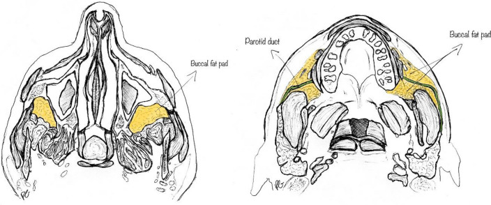
Background: The buccal space is an unusual location of malignancies. We report here the case of a woman with a melanoma metastasis in buccal fat pad, to evaluate the imaging features which might lead to the correct, although uncommon, diagnosis.
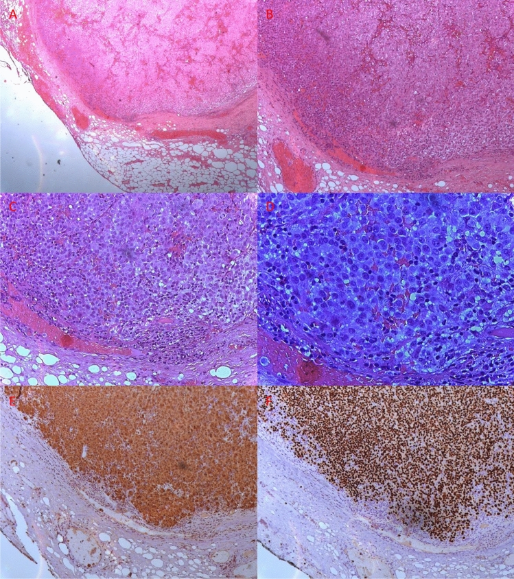
Case presentation: A 71-year-old woman presented with a painless visible swelling of the left cheek. MRI revealed the presence of a solid lesion located in the buccal fat pad with features suggestive of malignancy. It showed T1 hyperintensity and T2 hypointensity, and restriction of diffusion. Histological examination showed neoplastic cells compatible with melanoma.
Discussion: The lesion features (T1 hyperintensity and T2 hypointensity) initially lead our team to believe that there was a hemorrhagic component, possibly a residue of the biopsy. However, when associated with other malignancy features, such as low apparent diffusion coefficient (ADC) values and contrast enhancement, they should evoke the suspect of melanoma, provided that no biopsy was performed and no trauma occurred in the 3-7 days before.
Keywords: Buccal fat pad; MRI features; Melanoma; Melanoma metastasis.
Conflict of interest statement
The authors declare that they have no conflict of interest.
Figures


References
-
- Tart RP, Kotzur IM, Mancuso AA, Glantz MS, Mukherji SK. CT and MR imaging of the buccal space and buccal space masses. RadioGraphics. 1995;15(3):531–550. - PubMed
-
- Kurabayashi T, Ida M, Tetsumura A, Ohbayashi N, Yasumoto M, Sasaki T. MR imaging of benign and malignant lesions in the buccal space. Dentomaxillofac Radiol. 2002;31(6):344–349. - PubMed
-
- Gupta TD, Brasfield R. Metastatic melanoma. A clinicopathological study. Cancer. 1964;17(10):1323–1339. - PubMed
-
- Smoker WRK. Oral cavity. In: Som PM, Curtin HD, editors. Head and neck imaging. 3. St Louis, Mo: Mosby; 1996. pp. 488–544.