. 2020 Sep;51(9):2834-2843.
doi: 10.1161/STROKEAHA.120.029781. Epub 2020 Aug 10.

Rapamycin Induces an eNOS (Endothelial Nitric Oxide Synthase) Dependent Increase in Brain Collateral Perfusion in Wistar and Spontaneously Hypertensive Rats

Affiliations

Rapamycin Induces an eNOS (Endothelial Nitric Oxide Synthase) Dependent Increase in Brain Collateral Perfusion in Wistar and Spontaneously Hypertensive Rats

Daniel J Beard et al. Stroke. 2020 Sep.

Abstract

Background and purpose: Rapamycin is a clinically approved mammalian target of rapamycin inhibitor that has been shown to be neuroprotective in animal models of stroke. However, the mechanism of rapamycin-induced neuroprotection is still being explored. Our aims were to determine if rapamycin improved leptomeningeal collateral perfusion, to determine if this is through eNOS (endothelial nitric oxide synthase)-mediated vessel dilation and to determine if rapamycin increases immediate postreperfusion blood flow.

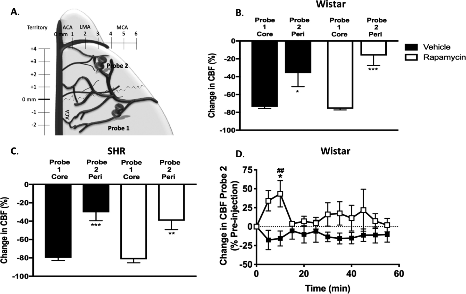
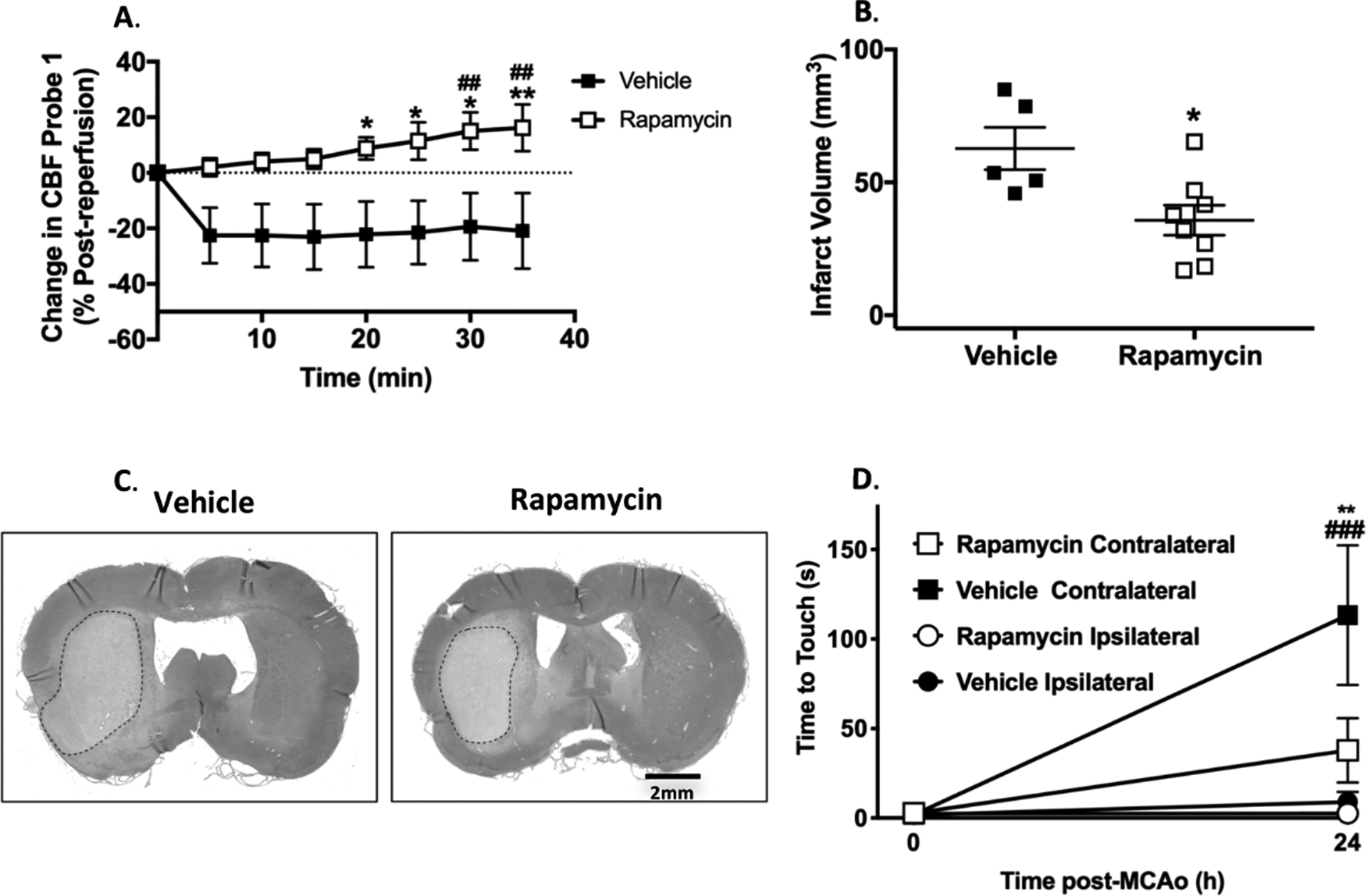
Methods: Wistar and spontaneously hypertensive rats (≈14 weeks old, n=22 and n=15, respectively) were subjected to ischemia by middle cerebral artery occlusion (90 and 120 minutes, respectively) with or without treatment with rapamycin at 30-minute poststroke. Changes in middle cerebral artery and collateral perfusion territories were measured by dual-site laser Doppler. Reactivity to rapamycin was studied using isolated and pressurized leptomeningeal anastomoses. Brain injury was measured histologically or with triphenyltetrazolium chloride staining.

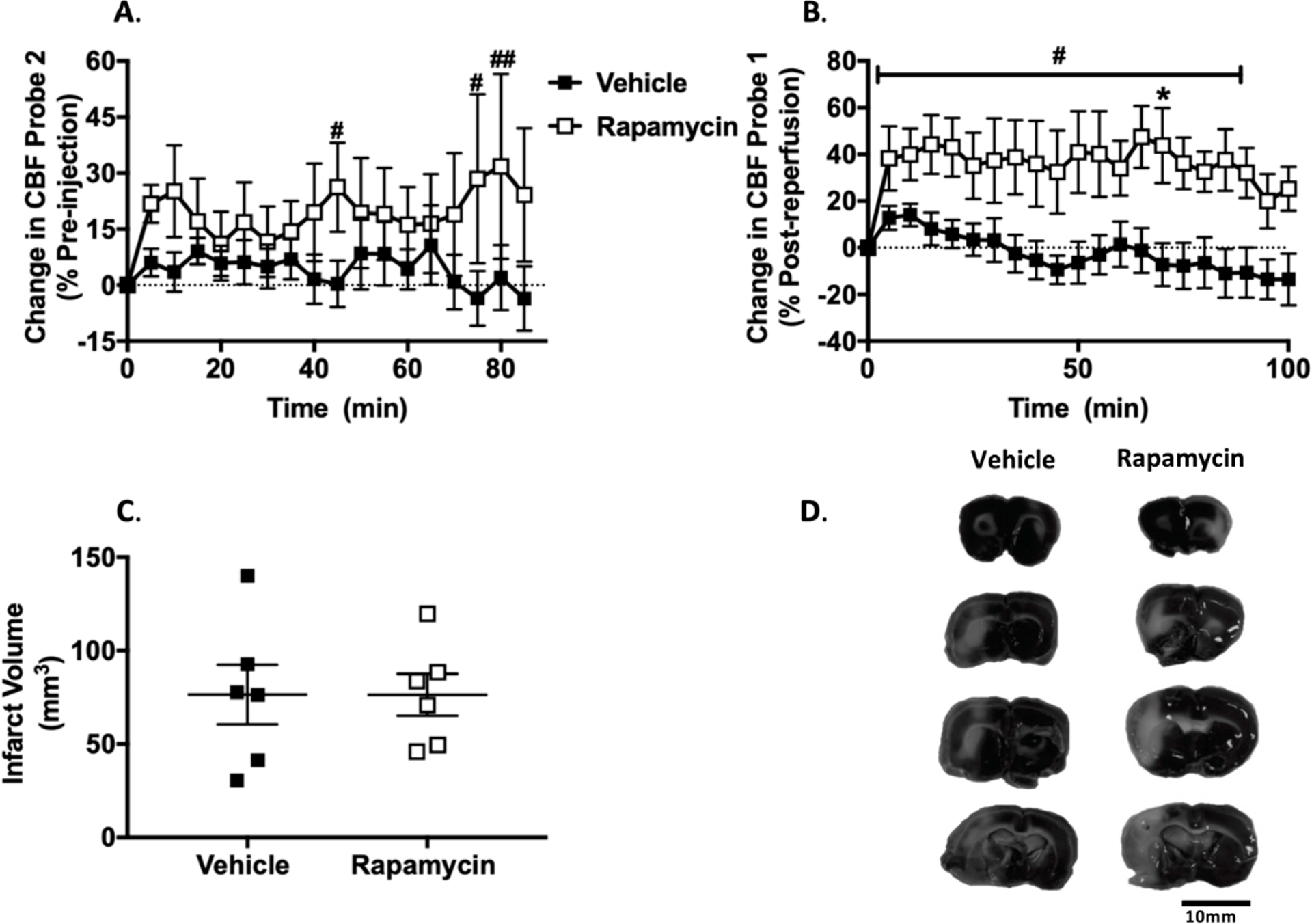
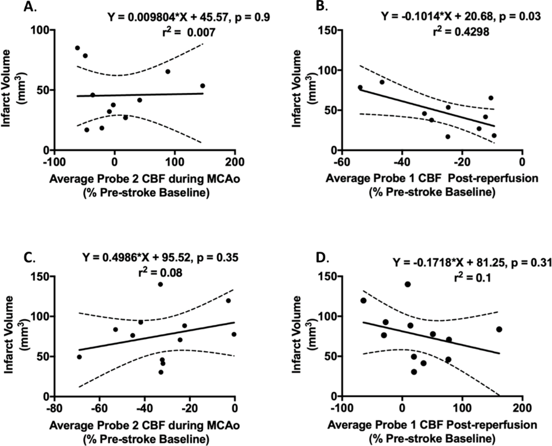
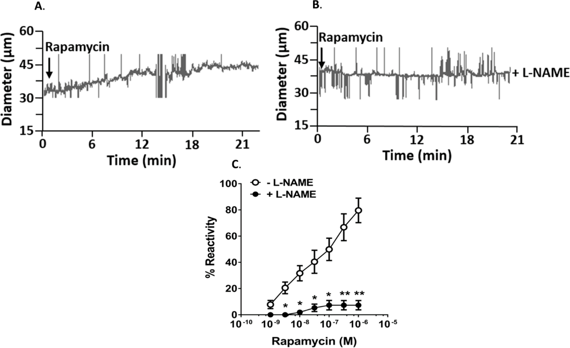
Results: In Wistar rats, rapamycin increased collateral perfusion (43±17%), increased reperfusion cerebral blood flow (16±8%) and significantly reduced infarct volume (35±6 versus 63±8 mm3, P<0.05). Rapamycin dilated leptomeningeal anastomoses by 80±9%, which was abolished by nitric oxide synthase inhibition. In spontaneously hypertensive rats, rapamycin increased collateral perfusion by 32±25%, reperfusion cerebral blood flow by 44±16%, without reducing acute infarct volume 2 hours postreperfusion. Reperfusion cerebral blood flow was a stronger predictor of brain damage than collateral perfusion in both Wistar and spontaneously hypertensive rats.

Conclusions: Rapamycin increased collateral perfusion and reperfusion cerebral blood flow in both Wistar and comorbid spontaneously hypertensive rats that appeared to be mediated by enhancing eNOS activation. These findings suggest that rapamycin may be an effective acute therapy for increasing collateral flow and as an adjunct therapy to thrombolysis or thrombectomy to improve reperfusion blood flow.

Keywords: dilation; hypertension; neuroprotection; reperfusion; stroke.


Conflict of interest statement

Disclosures

AMB is a senior medical science advisor and co-founder of Brainomix, a company that develops electronic ASPECTS (e-ASPECTS), an automated method to evaluate ASPECTS in stroke patients. All other authors declare no conflict of interest.

Figures

Figure 1.
Figure 1.. Multisite laser Doppler Flowmetry for measurement of collateral perfusion.
A. Schematic showing the location of the dual-site laser Doppler probes. B and C. Graphs comparing initial drop in cerebral blood flow (CBF) from baseline before filament insertion between MCA core (Probe 1) and peri-infarct (Probe 2) collateral CBF in Wistar rats and SHRs, respectively. Drop in collateral CBF (probe 2) was significantly less than that of the core MCA territory (probe 1) demonstrating that each probe was measuring different hemodynamic areas. *p < 0.05, **p < 0.01, *** p < 0.001 vs. probe 1 group, unpaired t-tests. D. Change in Leptomeningeal Anastomoses (LMA) CBF in Wistar rats calculated as % change from prior to treatment (30min after middle cerebral artery occlusion (MCAo). RM 2-Way ANOVA: F (1, 11) = 3.429, p = 0.09 for treatment, F (11, 121) = 0.5054, p > 0.05 for time, F (11, 121) = 1.463, p > 0.05 for interaction. *p < 0.05 vs. vehicle, Sidak’s post-test. ## p < 0.01 vs. pre-injection baseline in rapamycin group, Dunnett’s post-test.
Figure 2.
Figure 2.. Wistar rats reperfusion cerebral blood flow and stroke outcome analysis.
A. Change in middle cerebral artery (MCA) cerebral blood flow calculated as % change post- reperfusion (95 min post-MCAo). RM 2-Way ANOVA: F (1, 64) = 49.11, p < 0.0001 for treatment, F (7, 64) = 0.4979, p = 0.83 for time, F (7, 64) = 1.15, p = 0.35 for interaction. *p < 0.05, ** p < 0.01 vs. vehicle, Sidak’s post-test. ## p < 0.01 vs. post-reperfusion baseline in rapamycin group, Dunnett’s post-test. B. Infarct volume at 24 h post-MCAo. *p < 0.05 vs. vehicle, unpaired t-test. C. Representative histological hematoxylin and eosin stains for infarct volume. D. Time to touch in adhesive removal test at pre-MCAo baseline and 24 h after MCAo. RM 2-Way ANOVA: F (3, 22) = 6.253, p < 0.001 for treatment, F (1, 22) = 16.32, p < 0.01 for time, F (3, 22) = 6.375, p < 0.01 for interaction. **p < 0.01 rapamycin contralateral vs. vehicle contralateral, Tukey’s post-test. ### p < 0.001 vs. pre-MCAo baseline in vehicle group, Sidak’s post-test.
Figure 3.
Figure 3.. Spontaneously Hypertensive Rats (SHRs) studies.
A. Change in leptomeningeal anastomoses cerebral blood flow (CBF) calculated as % change from prior to treatment at 30min after middle cerebral artery occlusion (MCAo). RM 2-Way ANOVA: F (1, 10) = 1.403, p > 0.05 for treatment, F (17, 170) = 0.6585, p > 0.05 for time, F (17, 170) = 1.102, p > 0.05 for interaction. # p < 0.05, ## p < 0.01 vs. pre-injection baseline in rapamycin group, Dunnett’s post-test. B. Change in MCA CBF calculated as % change from reperfusion (125 min post-MCAo). RM 2-Way ANOVA: F (1, 10) = 8.433, p < 0.05 for treatment, F (20, 200) = 2.113, p < 0.01 for time, F (20, 200) = 1.202, p > 0.05 for interaction.* p < 0.05 vs. vehicle, RM 2-Way ANOVA, Sidak’s post-test. # p < 0.05 vs. post-reperfusion baseline in rapamycin group, RM 2-Way ANOVA, Dunnett’s post-test. C. Infarct volume at 4 h post-MCAo. D. Representative TTC stains.
Figure 4.
Figure 4.. Relationship between changes in leptomeningeal anastomoses (LMA) cerebral blood flow (CBF), reperfusion CBF and final infarct volume.
A-B. Wistar rats. A. Correlation between average LMA perfusion during middle cerebral artery occlusion (MCAo) and 24 hour infarct volume. There was a non-significant inverse correlation found between LMA blood flow and final infarct volume. B. Correlation between average reperfusion CBF and 24 hour infarct volume. There was a significant inverse correlation found between reperfusion CBF and infarct volume. C-D. SHRs. C. Correlation between average LMA perfusion during MCAo and 4 hour infarct volume. There was no significant correlation between LMA CBF and final infarct volume in SHRs. D. Correlation between Average reperfusion CBF and 4 hour infarct volume. There was no significant correlation between reperfusion CBF and infarct volume in SHRs.
Figure 5.
Figure 5.. Isolated leptomeningeal anastomoses (LMAs) from Wistar rats.
A and B. Representative tracing of changes in diameter of isolated and pressurized LMAs in response to rapamycin and rapamycin in the presence of L-NAME, respectively. C. Graph showing percent reactivity of isolated LMA to rapamycin in the absence and presence of L-NAME. Addition of rapamycin caused concentration-dependent vasodilation that was prevented by nitric oxide inhibition with L-NAME. * p < 0.05, **p < 0.001 vs. LMAs without L-NAME treatment.

References

    1. Benjamin EJ, Muntner P, Alonso A, Bittencourt MS, Callaway CW, Carson AP, Chamberlain AM, Chang AR, Cheng S, Das SR, et al. Heart disease and stroke statistics-2019 update a report from the american heart association. Circulation. 2019;139:E56–E528 - PubMed
    1. O’Collins VE, Macleod MR, Donnan GA, Horky LL, van der Worp BH, Howells DW. 1,026 experimental treatments in acute stroke. Ann Neurol. 2006;59:467–477 - PubMed
    1. Papadakis M, Hadley G, Xilouri M, Hoyte LC, Nagel S, McMenamin MM, Tsaknakis G, Watt SM, Drakesmith CW, Chen R, et al. Tsc1 (hamartin) confers neuroprotection against ischemia by inducing autophagy. Nat Med. 2013;19:351–357 - PMC - PubMed
    1. Huang J, Manning BD. The tsc1-tsc2 complex: A molecular switchboard controlling cell growth. Biochem J. 2008;412:179–190 - PMC - PubMed
    1. DeYoung MP, Horak P, Sofer A, Sgroi D, Ellisen LW. Hypoxia regulates tsc1/2-mtor signaling and tumor suppression through redd1-mediated 14-3-3 shuttling. Genes Dev. 2008;22:239–251 - PMC - PubMed

Publication types

MeSH terms