

HIV Impairs Alveolar Macrophage Function via MicroRNA-144-Induced Suppression of Nrf2
- PMID: 32773107
- PMCID: PMC7854972
- DOI: 10.1016/j.amjms.2020.07.026
HIV Impairs Alveolar Macrophage Function via MicroRNA-144-Induced Suppression of Nrf2
Abstract
Background: Despite anti-retroviral therapy, HIV-1 infection increases the risk of pneumonia and causes oxidative stress and defective alveolar macrophage (AM) immune function. We have previously determined that HIV-1 proteins inhibit antioxidant defenses and impair AM phagocytosis by suppressing nuclear factor (erythroid-derived 2)-like 2 (Nrf2). Given its known effects on Nrf2, we hypothesize miR-144 mediates the HIV-1 induced suppression of Nrf2.
Methods: Primary AMs isolated from HIV-1 transgenic (HIV-1 Tg) rats and wild type littermates (WT) as well as human monocyte-derived macrophages (MDMs) infected ex vivo with HIV-1 were used. We modulated miR-144 expression using a miR-144 mimic or an inhibitor to assay its effects on Nrf2/ARE activity and AM functions in vitro and in vivo.
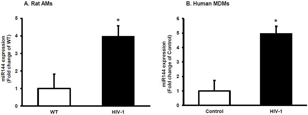
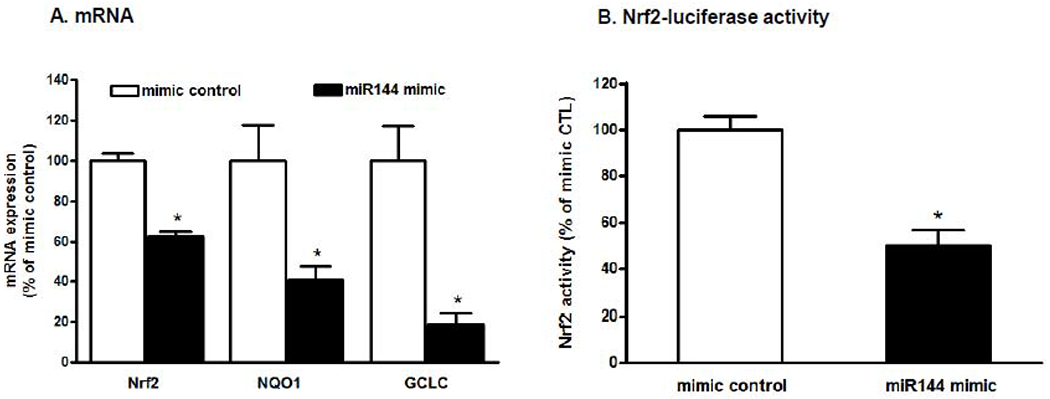
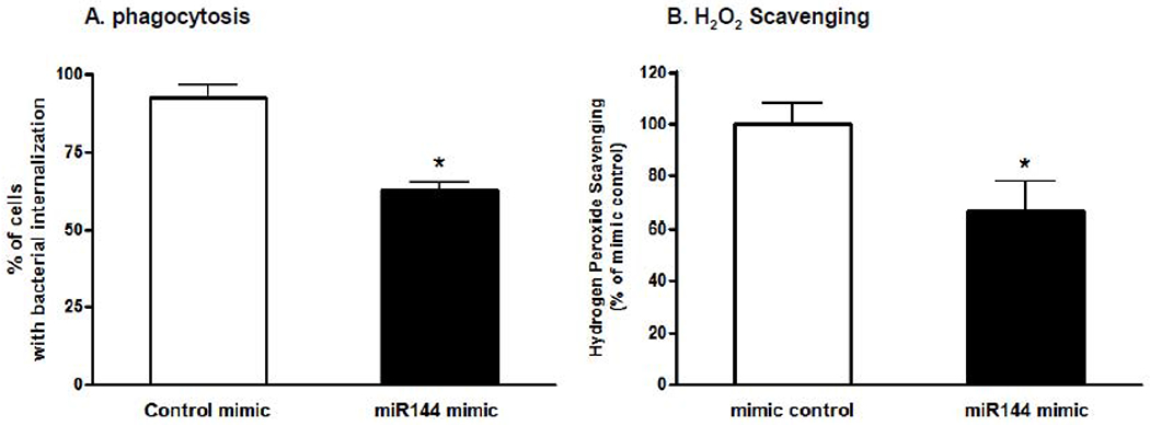
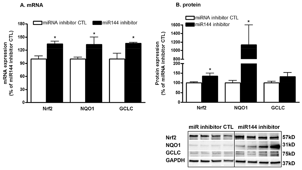
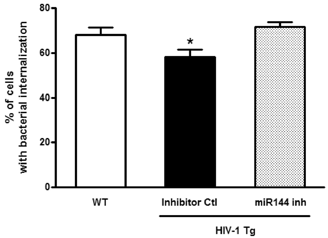
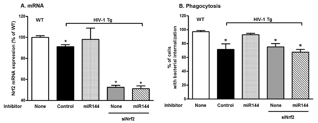
Results: MiR-144 expression was increased in AMs from HIV-1 Tg rats and in HIV-1-infected human MDMs compared to cells from WT rats and non-infected human MDMs, respectively. Increasing miR-144 with a miR-144 mimic inhibited the expression of Nrf2 and its downstream effectors in WT rat macrophages and consequently impaired their bacterial phagocytic capacity and H2O2 scavenging ability. These effects on Nrf2 expression and AM function were reversed by antagonizing miR-144 ex vivo or in the airways of HIV-1 Tg rats in vivo, but this protection was abrogated by silencing Nrf2 expression.
Conclusions: Our results suggest that inhibiting miR-144 or interfering with its deleterious effects on Nrf2 attenuates HIV-1-mediated AM immune dysfunction and improves lung health in individuals with HIV.
Keywords: Alveolar macrophage; HIV-1 transgenic rat; MIR-144; Nrf2.
Copyright © 2020 Southern Society for Clinical Investigation. Published by Elsevier Inc. All rights reserved.
Conflict of interest statement
Figures
Similar articles
-
HIV-1 decreases Nrf2/ARE activity and phagocytic function in alveolar macrophages.J Leukoc Biol. 2017 Aug;102(2):517-525. doi: 10.1189/jlb.4A0616-282RR. Epub 2017 May 26. J Leukoc Biol. 2017. PMID: 28550120 Free PMC article.
-
MiR-144 mediates Nrf2 inhibition and alveolar epithelial dysfunction in HIV-1 transgenic rats.Am J Physiol Cell Physiol. 2019 Aug 1;317(2):C390-C397. doi: 10.1152/ajpcell.00038.2019. Epub 2019 May 15. Am J Physiol Cell Physiol. 2019. PMID: 31091144 Free PMC article.
-
Nrf2 regulates PU.1 expression and activity in the alveolar macrophage.Am J Physiol Lung Cell Mol Physiol. 2015 May 15;308(10):L1086-93. doi: 10.1152/ajplung.00355.2014. Epub 2015 Apr 3. Am J Physiol Lung Cell Mol Physiol. 2015. PMID: 25840997 Free PMC article.
-
Crosstalk between NRF2 and Dicer through metastasis regulating MicroRNAs; mir-34a, mir-200 family and mir-103/107 family.Arch Biochem Biophys. 2020 Jun 15;686:108326. doi: 10.1016/j.abb.2020.108326. Epub 2020 Mar 3. Arch Biochem Biophys. 2020. PMID: 32142889 Review.
-
Bacterial Infections in Patients Living with HIV.Results Probl Cell Differ. 2024;73:537-549. doi: 10.1007/978-3-031-62036-2_21. Results Probl Cell Differ. 2024. PMID: 39242392 Free PMC article. Review.
Cited by
-
Mitochondria as a Cellular Hub in Infection and Inflammation.Int J Mol Sci. 2021 Oct 20;22(21):11338. doi: 10.3390/ijms222111338. Int J Mol Sci. 2021. PMID: 34768767 Free PMC article. Review.
-
Keap1 recognizes EIAV early accessory protein Rev to promote antiviral defense.PLoS Pathog. 2022 Feb 9;18(2):e1009986. doi: 10.1371/journal.ppat.1009986. eCollection 2022 Feb. PLoS Pathog. 2022. PMID: 35139135 Free PMC article.
-
MicroRNAs: Small but Key Players in Viral Infections and Immune Responses to Viral Pathogens.Biology (Basel). 2023 Oct 14;12(10):1334. doi: 10.3390/biology12101334. Biology (Basel). 2023. PMID: 37887044 Free PMC article. Review.
-
Research progress on miRNAs function in the interaction between human infectious viruses and hosts: A review.Biomol Biomed. 2024 Oct 17;24(6):1452-1462. doi: 10.17305/bb.2024.10821. Biomol Biomed. 2024. PMID: 39101759 Free PMC article. Review.
-
Circadian-Coupled Genes Expression and Regulation in HIV-Associated Chronic Obstructive Pulmonary Disease (COPD) and Lung Comorbidities.Int J Mol Sci. 2023 May 23;24(11):9140. doi: 10.3390/ijms24119140. Int J Mol Sci. 2023. PMID: 37298092 Free PMC article. Review.
References
-
- UNAIDS. Global HIV & AIDS statistics - 2019 fact sheet. 2019.
-
- Afessa B, Green W, Chiao J, et al. Pulmonary complications of HIV infection: autopsy findings. Chest 1998;113:1225–9. - PubMed
-
- Hung CC, Chang SC. Impact of highly active antiretroviral therapy on incidence and management of human immunodeficiency virus-related opportunistic infections. J Antimicrob Chemother 2004;54:849–53. - PubMed
Publication types
MeSH terms
Substances
Grants and funding
LinkOut - more resources
Full Text Sources
Other Literature Sources
Medical
Miscellaneous
