. 2019 Aug;58(8):421-437.
doi: 10.1007/s00397-019-01142-2. Epub 2019 Apr 25.

Characterizing the dynamic rheology in the pericellular region by human mesenchymal stem cell re-engineering in PEG-peptide hydrogel scaffolds

Affiliations

Characterizing the dynamic rheology in the pericellular region by human mesenchymal stem cell re-engineering in PEG-peptide hydrogel scaffolds

Maryam Daviran et al. Rheol Acta. 2019 Aug.

Abstract

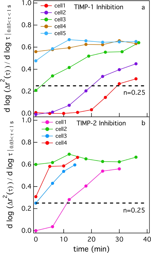
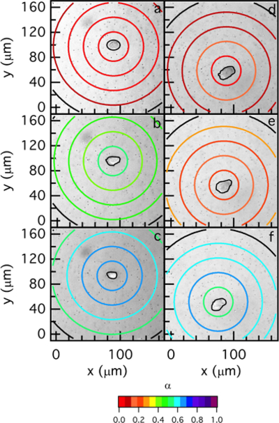
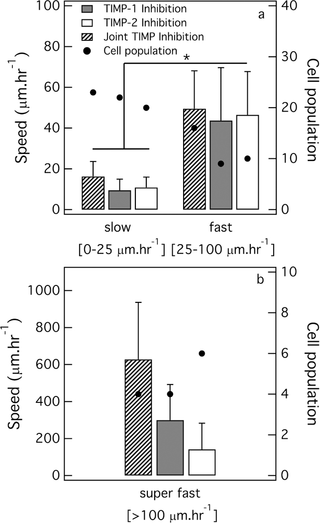
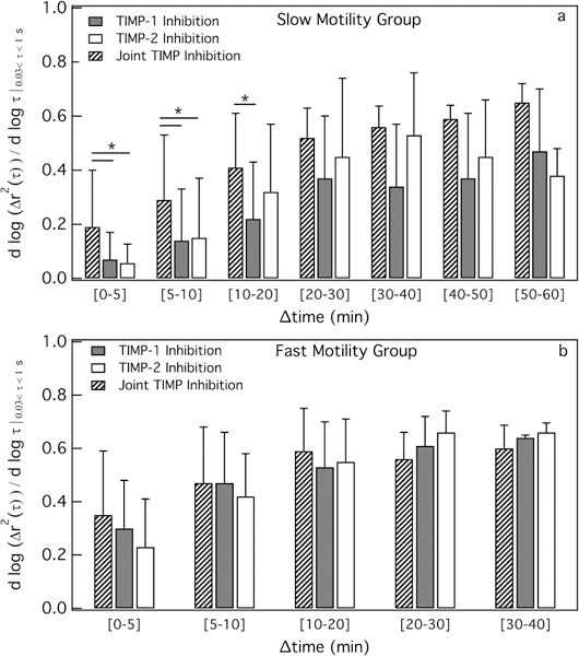
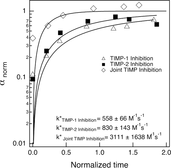
During wound healing, human mesenchymal stem cells (hMSCs) migrate to injuries to regulate inflammation and coordinate tissue regeneration. To enable migration, hMSCs re-engineer the extracellular matrix rheology. Our work determines the correlation between cell engineered rheology and motility. We encapsulate hMSCs in a cell-degradable peptide-polymeric hydrogel and characterize the change in rheological properties in the pericellular region using multiple particle tracking microrheology. Previous studies determined that pericellular rheology is correlated with motility. Additionally, hMSCs re-engineer their microenvironment by regulating cell-secreted enzyme, matrix metallopro-teinases (MMPs), activity by also secreting their inhibitors, tissue inhibitors of metalloproteinases (TIMPs). We independently inhibit TIMPs and measure two different degradation profiles, reaction-diffusion and reverse reaction-diffusion. These profiles are correlated with cell spreading, speed and motility type. We model scaffold degradation using Michaelis-Menten kinetics, finding a decrease in kinetics between joint and independent TIMP inhibition. hMSCs ability to regulate microenvironmental remodeling and motility could be exploited in design of new materials that deliver hMSCs to wounds to enhance healing.

Keywords: Michaelis-Menten kinetics; Multiple particle tracking microrheology; cellular degradation; matrix metalloproteinases; polymeric hydrogel scaffold; tissue inhibitor of metalloproteinases.


Figures

Fig. 1
Fig. 1
Changes in the logarithmic slope of mean-squared displacement,α=dlogΔr2(τ)dlogτ over time for (a) TIMP-1 and (b) TIMP-2 inhibited hMSCs. The critical relaxation exponent, n, in both graphs (a, b) is show with dashed lines. n quantifies the transition from a gel to a sol.
Fig. 2
Fig. 2
Spatial and temporal degradation profiles around two different TIMP-1 inhibited hMSCs. The left column is MPT data around an hMSC which has a reaction-diffusion degradation profile and the right column is MPT data for an hMSC which has a reverse reaction-diffusion degradation profile. MPT data are collected at (a) 0, (b) 18, (c) 31, (d) 0, (e) 12 and (f) 42 min. t = 0 min is the beginning of data acquisition. The color of the each ring is α=dlogΔr2(τ)dlogτ, which represents the state of the material in the hydrogel.
Fig. 3
Fig. 3
Average hMSC speed and hMSC population for joint TIMP, TIMP-1 inhibited and TIMP−2 inhibited hMSCs in the (a) slow, fast and (b) super fast motility groups. The average cell speed for individual TIMP inhibition and joint TIMP inhibition are not significantly significant but are significant between the slow and fast motility groups for the same cell treatment (*p < 0.05).
Fig. 4
Fig. 4
The change in the average logarithmic slope of mean-squared displacement,α=dlogΔr2(τ)dlogτ at each time interval for joint and independently inhibited hMSCs with different migration speeds. The change in αavg for each time interval for the a) slow and b) fast motility groups. In the slow motility group, αavg for joint inhibited hMSCs is significantly (*p < 0.05) higher than individually inhibited hMSCs until 20 min.
Fig. 5
Fig. 5
Experimental data and Michaelis-Menten enzymatic kinetic model for joint and independently inhibited hMSCs.αnorm=ααmaxαmin is plotted versus tnorm=ttavg. The experimental data is fit with Equation 5 to determine k* for each experiment. Joint inhibited hMSCs have an order of magnitude higher k* value because all MMPs are actively degrading the scaffold. In independently inhibited hMSC experiments some MMPs are inhibited by the remaining active TIMP.
Fig. 6
Fig. 6
Cell distribution and morphology in a hydrogel scaffold 18–48 hr after encapsulation. a) Quantitative calculation of cell percentage at different z positions indicates that both untreated and joint TIMP inhibited hMSCs are concentrated in the bottom of the hydrogel. b) Cells are distributed approximately equally in the edges and center of the hydrogel in joint TIMP inhibited and untreated experiments. d) 3D distribution of untreated hMSCs in the hydrogel scaffold and e) joint TIMP inhibited hMSCs in the hydrogel scaffold, scale bar = 30 μm. f) Elliptical form factor for each cell calculated by dividing cell length by its perpendicular width. Untreated hMSCs are significantly (*p < 0.05) more spread in the hydrogel.

Similar articles

Cited by

References

    1. abcam (2018) buffer and stock solutions for western blot. https://wwwabcamcom/protocols/buffer-and-stock-solutions-for-western-blot
    1. Adolf D, Martin JE (1990) Time-cure superposition during crosslinking. Macromolecules 23:3700–3704
    1. Aimetti AA, Machen AJ, Anseth KS (2009) Poly(ethylene glycol) hydrogels formed by thiol-ene photopolymerization for enzyme-responsive protein delivery. Biomaterials 30:6048–6054 - PMC - PubMed
    1. Anderson SB, Lin CC, Kuntzler DV, Anseth KS (2011) The performance of human mesenchymal stem cells encapsulated in cell-degradable polymer-peptide hydrogels. Biomaterials 32:3564–3574 - PMC - PubMed
    1. Bassi EJ, Candido de ALmeida D, Moraes-Vieira PMM, Camara NOS (2012) Exploring the role of soluble factors associated with immune regulatory properties of mesenchymal stem cells. Stem Cell Rev and Rep 8:329–342 - PubMed

LinkOut - more resources