Review
. 2020 Aug;37(3):227-236.
doi: 10.1055/s-0040-1713440. Epub 2020 Jul 31.

Lymphatic Anatomy and Physiology

Affiliations
Review

Lymphatic Anatomy and Physiology

Abhishek K Goswami et al. Semin Intervent Radiol. 2020 Aug.

Abstract

Lymphatics have long been overshadowed by the remainder of the circulatory system. Historically, lymphatics were difficult to study because of their small and indistinct vessels, colorless fluid contents, and limited effective interventions. However, the past several decades have brought increased funding, advanced imaging technologies, and novel interventional techniques to the field. Understanding the history of lymphatic anatomy and physiology is vital to further realize the role lymphatics play in most major disease pathologies and innovate interventional solutions for them.

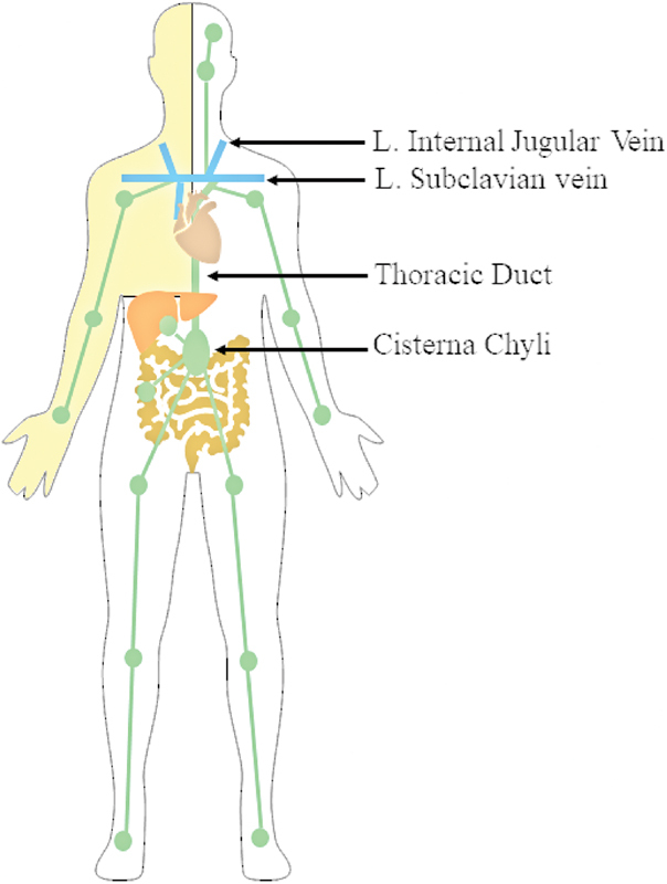
Keywords: anatomy; cisterna chyli; interventional radiology; lymph node; lymphatic; thoracic duct.


Conflict of interest statement

Conflict of Interest None declared.

Figures

Fig. 1
Fig. 1
Overview of lymphatic flow in the body. The lower extremities, left upper extremity, left head, left hemithorax, and subdiaphragmatic organs drain into the cisterna chyli and transit through the thoracic duct emptying into the left venous angle, near the junction of the left internal jugular and subclavian veins. The right upper extremity, head, and hemithorax (drainage pattern shaded yellow) drain to the right lymphatic duct, near the junction between the right internal jugular and subclavian veins.
Fig. 2
Fig. 2
Diagram of lymphatic microanatomy. Initial lymphatic vessels converge into collecting lymphatic vessels, which flow into lymph nodes. Initial lymphatics have leaky flap junctions allowing fluid to enter while collecting lymphatics have tight junctions surrounded by smooth muscle to propel lymph forward.
Fig. 3
Fig. 3
Volume-rendered CT lymphangiography in coronal ( a ) and sagittal ( b ) planes shows coalescing of lymphatic vessels along the pelvic sidewalls to the retroperitoneum overlying the spine.
Fig. 4
Fig. 4
Radiograph of central lymphatics during lymphangiography reveals a saccular structure at the bottom of the image (black arrow) representing the cisterna chyli, which gives rise to a single main thoracic duct (white arrow), which crosses from right to left as it ascends toward the neck.
Fig. 5
Fig. 5
Radiograph of the terminal thoracic duct during lymphangiography. The terminal thoracic duct (white arrow) ascends above the left clavicle and has two valves at its communication with the left venous angle (black arrow).
Fig. 6
Fig. 6
Pedal lymphangiography of the left leg. ( a ) A 30-gauge needle (white arrowhead) opacifying a dermal foot lymphatic vessel (white arrow). As ethiodized oil ascends into the ( b ) lower leg, ( c ) knee, and ( d ) thigh, additional channels begin to fill (white arrowheads). All the lymphatic vessels are similar in size. No lymph nodes were visualized from the foot to the thigh. At the level of the pelvis ( e ), the lymphatic vessels (black arrowheads) converge into inguinal lymph nodes, first seen central to the lesser trochanter, which transmit the dye through multiple lymphatic vessels (white arrow). ( f ) Interspersed lymph nodes and lymphatic vessels (white arrowhead) are present from the inguinal area, along the pelvic sidewall and into the lumbar lymphatic chain (white arrow).
Fig. 7
Fig. 7
Schematic showing intestinal microvillus. Each microvillus has an arteriovenous capillary network and a lacteal, with the lacteal absorbing long-chain fatty acids, which are processed into chylomicrons before draining to mesenteric lymph nodes and subsequently the thoracic duct.
Fig. 8
Fig. 8
Diagram of a hepatic sinusoid, depicting origin of hepatic lymph. Blood enters the sinusoid from branches of the portal vein and of the hepatic artery, which allows mixing. Fluid extravasates into the space of “Disse” to flow retrograde, entering initial lymphatic microvessels surrounding the portal vein.
Fig. 9
Fig. 9
Hepatic lymphangiography and cholangiography. A 22-gauge needle was passed into the liver and contrast was injected while it was withdrawn, opacifying ( a ) a peripheral lymphatic vessel (black arrow) and biliary radical (white arrow). ( b ) The biliary tree (white arrow) becomes larger as it centralizes, while the lymphatics (black arrow) do not markedly enlarge in size and bifurcate. ( c ) The 22-gauge needle was repositioned centrally in the hepatic hilum and opacified the lymphatics (black arrow), which are tortuous, redundant, and extend to the hepatoduodenal ligament, along the portal vein. Cholecystectomy clips are present at the bottom of the image and a microwire is present in the left hepatic duct and central biliary tree.

References

    1. Natale G, Bocci G, Ribatti D. Scholars and scientists in the history of the lymphatic system. J Anat. 2017;231(03):417–429. - PMC - PubMed
    1. Breslin J W, Yang Y, Scallan J P, Sweat R S, Adderley S P, Murfee W L. Lymphatic vessel network structure and physiology. Compr Physiol. 2018;9(01):207–299. - PMC - PubMed
    1. Mortimer P S, Rockson S G. New developments in clinical aspects of lymphatic disease. J Clin Invest. 2014;124(03):915–921. - PMC - PubMed
    1. Renkin E M.Some consequences of capillary permeability to macromolecules: Starlings hypothesis reconsideredAm J Physiol Heart Circ Physiol1986. 250(05): - PubMed
    1. Baluk P, Fuxe J, Hashizume H et al.Functionally specialized junctions between endothelial cells of lymphatic vessels. J Exp Med. 2007;204(10):2349–2362. - PMC - PubMed