Case Reports
. 2020 May-Jun;24(3):276-279.
doi: 10.4103/jisp.jisp_459_19. Epub 2020 Jan 27.

An aggressive pyogenic granuloma masquerading as a vascular neoplasm

Affiliations
Case Reports

An aggressive pyogenic granuloma masquerading as a vascular neoplasm

Shravya Jaganath Shetty et al. J Indian Soc Periodontol. 2020 May-Jun.

Abstract

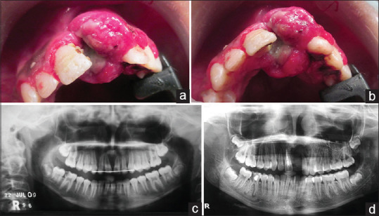
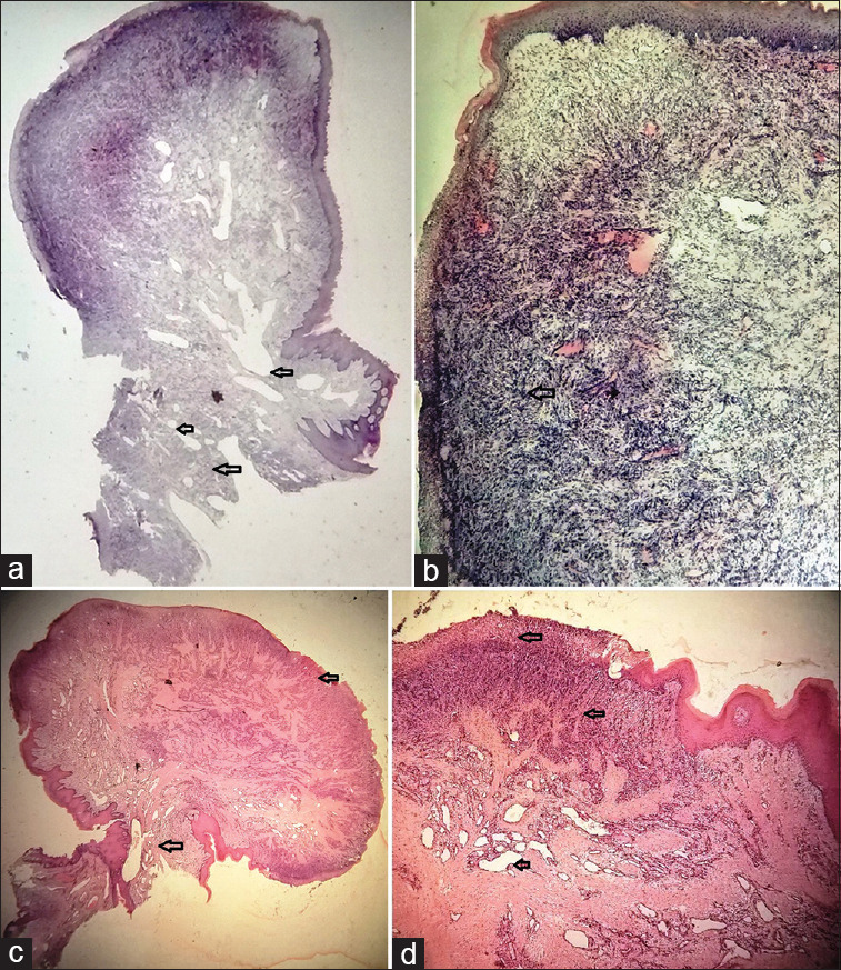
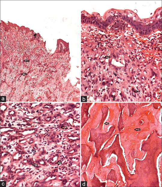
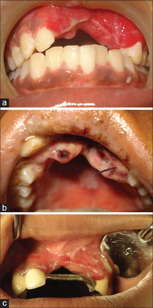
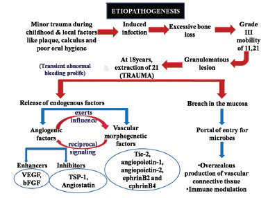
Pyogenic granuloma (PG) is a benign lesion, with a female predilection, commonly associated with local irritation or trauma. We report an unusual, destructive case of PG, displaying excessive loss of blood and destruction of alveolar bone leading to the loss of maxillary anterior teeth in an 18-year-old female, compromising function and esthetics. The incisional and excisional biopsy specimen of this recurrent lesion obtained during a 5-year span was studied, which revealed an increase in vascularity and extensive proliferation of endothelial cells admixed with varying degree of inflammatory cell infiltrate. The clinical, radiographic, and histopathological diagnostic tools enabled to precisely diagnose the lesion as an aggressive variant of PG, distinguishing it from other vascular neoplasms. No recurrence has been noted during a 5-year follow-up. The clinicians should be aware of the aggressive and destructive clinical behavior of PG to avoid the inadvertent treatment of this reactive lesion.

Keywords: Aggressive; pyogenic granuloma; recurrent; vascular neoplasm.


Conflict of interest statement

There are no conflicts of interest.

Figures

Figure 1
Figure 1
(a and b) Show a diffuse bright red lesion with a lobulated surface is noted in the labial and palatal aspect extending from 11 to 23 with ill-defined margins; (c) In the initial presentation, the orthopantomography shows a well-defined radiolucency in the interdental region of 11, 21; (d) In the second recurrence, the orthopantomography shows ill-defined radiolucency in the interradicular area extending into the alveolus and palatal region from 11 to 22
Figure 2
Figure 2
(a and b) Initial presentation showing a pedunculated mass with an ulcerated epithelium and stroma is chronically inflamed, cellular, vascular with endothelial cell proliferation. (H and E, low-magnification views ×1.25 and ×10); (c and d) first recurrence showing a pedunculated growth with surface ulceration and the vascularity extending till the stalk of the lesion. Note the increase in the size and the vascularity of the lesion. (H and E, low-magnification views ×1.25 and ×10)
Figure 3
Figure 3
Second recurrence- (a-c) show the discontinuous oral epithelium with the adjacent stroma showing numerous capillaries lined with plump endothelial cell proliferation and lymphocytic infiltration; (a: H and E stains, low-magnification view ×10; b and c: H and E, high-magnification views ×40); (d) The decalcified section of the bony trabeculae with dilated blood vessels. (H and E, high-magnification views ×40)
Figure 4
Figure 4
(a) Followups showing wellhealed lesions displaying loss of alveolar bone and soft tissue; (b) and erythematous lesion in the second followup. (c) The missing teeth are replaced with a fixed prosthesis
Figure 5
Figure 5
The possible etiopathogenesis of the present case wherein, trauma, local and systemic factors, and microorganisms play a vital role. (VEGF – Vascular endothelial growth factor; bFGF – Basic fibroblast growth factor; TSP-1 – Thrombospondin 1; Tie-2 – Tyrosine kinase with immunoglobulin-like and EGF-like domains-2)

References

    1. Mastammanavar D, Hunasgi S, Koneru A, Vanishree M, Surekha R, Vardendra M. Aggressive pyogenic granuloma: A case report. Int J Oral Maxillofac Pathol. 2014;5:29–32.
    1. Mohan M, Karikal A, Bhat S, Padmaja A, Thilak G. Aggressive pyogenic granuloma causing bone erosion. A case report. Healtalk. 2012;4:12–4.
    1. Martins-Filho PR, Piva MR, Da Silva LC, Reinheimer DM, Santos TS. Aggressive pregnancy tumor (pyogenic granuloma) with extensive alveolar bone loss mimicking a malignant tumor: Case report and review of literature. Int J Morphol. 2011;29:164–167.
    1. Singh RK, Kaushal A, Kumar R, Pandey RK. Profusely bleeding oral pyogenic granuloma in a teenage girl. BMJ Case Rep. 2013;2013:bcr2013008583. - PMC - PubMed
    1. Panseriya BJ, Hungund S. Pyogenic granuloma associated with periodontal abscess and bone loss – A rare case report. Contemp Clin Dent. 2011;2:240–4. - PMC - PubMed

Publication types