. 2020 Jul 3:14:189.
doi: 10.3389/fncel.2020.00189. eCollection 2020.

Potential Involvement of Adiponectin Signaling in Regulating Physical Exercise-Elicited Hippocampal Neurogenesis and Dendritic Morphology in Stressed Mice

Affiliations

Potential Involvement of Adiponectin Signaling in Regulating Physical Exercise-Elicited Hippocampal Neurogenesis and Dendritic Morphology in Stressed Mice

Pingjie Wang et al. Front Cell Neurosci. .

Abstract

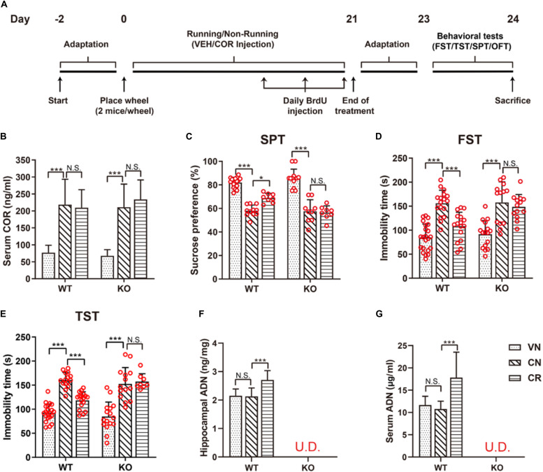
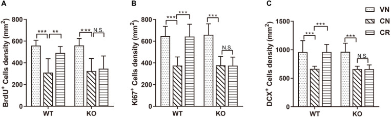
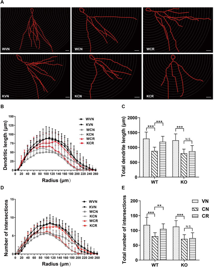
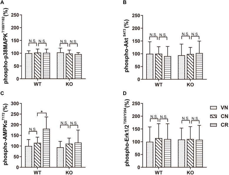
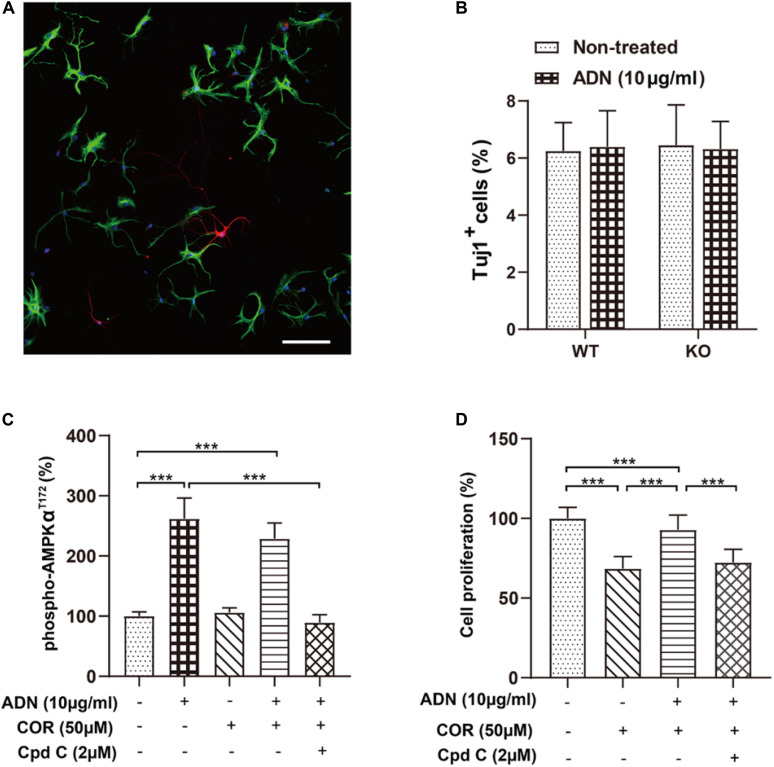
Adiponectin, a cytokine secreted by mature adipocytes, proves to be neuroprotective. We have previously reported that running triggers adiponectin up-regulation which subsequently promotes generation of hippocampal neurons and thereby alleviates depression-like behaviors in non-stressed mice. However, under the stressing condition, whether adiponectin could still exert antidepressant-like effects following exercise remained unexplored. In this study, by means of repeated corticosterone injections to mimic stress insult and voluntary wheel running as physical exercise intervention, we examined whether exercise-elicited antidepressive effects might involve adiponectin's regulation on hippocampal neurogenesis and dendritic plasticity in stressed mice. Here we show that repeated injections of corticosterone inhibited hippocampal neurogenesis and impaired dendritic morphology of neurons in the dentate gyrus of both wild-type and adiponectin-knockout mice comparably, which subsequently evoked depression-like behaviors. Voluntary wheel running attenuated corticosterone-suppressed neurogenesis and enhanced dendritic plasticity in the hippocampus, ultimately reducing depression-like behaviors in wild-type, but not adiponectin-knockout mice. We further demonstrate that such proneurogenic effects were potentially achieved through activation of the AMP-dependent kinase (AMPK) pathway. Our study provides the first evidence that adiponectin signaling is essential for physical exercise-triggered effects on stress-elicited depression by retaining the normal proliferation of neural progenitors and dendritic morphology of neurons in the hippocampal dentate gyrus, which may depend on activation of the AMPK pathway.

Keywords: adiponectin; dendritic plasticity; dentate gyrus; depression; hippocampal neurogenesis; voluntary exercise.


Figures

FIGURE 1
FIGURE 1
Voluntary exercise is insufficient to reduce depression-like behaviors in ADN-KO mice under stress. (A) Experimental timeline for animal treatments. (B) Changes of the serum COR level following administration of COR with or without running. n = 10–11 mice/group. (C–E) Assessments of behavioral despairs after different treatments. There was no main difference of genotype in COR-induced depressive behaviors. However, voluntary exercise substantially raised sucrose preference in the SPT (C; n = 8–18 mice/group), decreased immobility time in the FST (D; n = 13–22 mice/group) and the TST (E; n = 8–21 mice/group) in COR-treated WT mice; these effects were diminished in ADN-deficient mice. (F,G) COR injections did not affect the hippocampal (F; n = 8 mice/group) or serum ADN level (G; n = 8 mice/group) in WT non-runners, while the 3-week voluntary running raised both. ADN was undetected in KO mice with the designated treatments. Data are shown as means ± SD. *p < 0.05, ***p < 0.001 by two-way ANOVA and Tukey’s post hoc test. N.S., non-significant; U.D., undetected; FST, forced swim test; TST, tail suspension test; SPT, sucrose preference test; OFT, open field test; COR, corticosterone; ADN, adiponectin.
FIGURE 2
FIGURE 2
Adiponectin deficiency attenuates exercise-enhanced neurogenesis under stress. (A–C) COR treatment significantly lowered the number of BrdU+ newborn cells (A; n = 11–12 mice/group), Ki67+ proliferating cells (B; n = 9–15 mice/group) and DCX+ immature neurons (C; n = 9–15 mice/group) in both WT and ADN-KO mice. Voluntary exercise enhanced hippocampal neurogenesis by increasing all these three subsets of cells in COR-treated WT (WCR), but not ADN-null mice (KCR). Data are shown as means ± SD. **p < 0.01, ***p < 0.001 by two-way ANOVA and Tukey’s post hoc test. N.S., non-significant; BrdU, bromodeoxyuridine; Ki67, cell proliferation-associated nuclear antigen; DCX, doublecortin.
FIGURE 3
FIGURE 3
Adiponectin is required for exercise to mitigate corticosterone-elicited impairment of dendritic reorganization. (A) The typical morphology of neurons was reconstructed with Neurolucida software. Scale bars: 20 μm. (B,C) COR treatment remarkably decreased the dendritic length, as illustrated by the distribution plot of dendritic length (B; n = 12–17 neurons/group) and the average of total dendritic length (C; n = 12–17 neurons/group). (D–E) The number of intersections also declined after COR treatments by Sholl analysis, as evidenced by the distribution plot of the number of intersections (D; n = 12–17 neurons/group) and the average of total intersections (E; n = 12–17 neurons/group). Notably, exercise significantly increased the dendritic length and the number of intersections in WCR rather than KCR. Data are shown as means ± SD. **p < 0.01, ***p < 0.001 by two-way ANOVA and Tukey’s post hoc test. N.S., non-significant.
FIGURE 4
FIGURE 4
Voluntary exercise selectively activates the AMPK pathway through adiponectin. Levels of phospho-p38MAPKT180/Y182, phospho-AKTS473, phospho-AMPKT172, and phospho-Erk1/2T202/Y204 following various treatments were quantified by ELISA. (A–D) In both strains, COR and exercise did not affect the expression of phospho-p38MAPKT180/Y182 (A), phospho-AKTS473 (B), or phospho-Erk1/2T202/Y204 (D). However, exercise significantly raised the phospho-AMPKT172 level in stressed WT, but not ADN-KO mice (C). Data are shown as means ± SD. n = 10 mice/group. *p < 0.05 by two-way ANOVA and Tukey’s post hoc test. N.S., non-significant; MAPK, mitogen-activated protein kinase; AKT, protein kinase B; AMPK, AMP-activated protein kinase; Erk, extracellular signal-regulated kinase.
FIGURE 5
FIGURE 5
Inhibition of the AMPK pathway diminishes adiponectin-enhanced cell proliferation in vitro. (A) The representative image showing that NPCs grown on coverslips after a 6-day culture in the induction medium differentiated into the Tuj1+ neuronal cells (red) and the GFAP+ glial cells (green) with the corresponding diverse morphologies. Scale bar: 100 μm. (B) Adding ADN into the medium to a final concentration of 10 μg/ml did not affect the percentage of induced neuronal cells in either WT or KO NPCs; the basal neuronal differentiation was unaltered after ADN KO. N = 6 coverslips/group from 3 independent experiments. (C) The phosphorylation of AMPK at the T172 site after application of ADN, COR and Cpd C. COR did not affect the level of phospho-AMPKT172, whereas Cpd C essentially abolished adiponectin-induced enhancement of AMPK phosphorylation. N = 4 independent experiments. (D) Cell proliferation following ADN, COR, and Cpd C treatments. Administration of COR significantly impaired neuronal proliferation which could be reversed by adiponectin addition, whereas inhibition of AMPK phosphorylation by Cpd C potently abolished the aforementioned proliferating effect of adiponectin. N = 3 independent experiments. Data are shown as means ± SD. ***p < 0.001 by one-way ANOVA and Tukey’s post hoc test. Tuj1, Neuronal Class III β-Tubulin (neuronal marker); GFAP, glial fibrillary acidic protein (glial marker); COR, corticosterone; ADN, adiponectin; Cpd C, Compound C (AMPK antagonist).

References

    1. Abdollahi A., LeBouthillier D. M., Najafi M., Asmundson G. J. G., Hosseinian S., Shahidi S., et al. (2017). Effect of exercise augmentation of cognitive behavioural therapy for the treatment of suicidal ideation and depression. J. Affect. Disord. 219 58–63. 10.1016/j.jad.2017.05.012 - DOI - PubMed
    1. Adlard P. A., Perreau V. M., Engesser-Cesar C., Cotman C. W. (2004). The timecourse of induction of brain-derived neurotrophic factor mRNA and protein in the rat hippocampus following voluntary exercise. Neurosci. Lett. 363 43–48. 10.1016/j.neulet.2004.03.058 - DOI - PubMed
    1. Agudelo L. Z., Femenia T., Orhan F., Porsmyr-Palmertz M., Goiny M., Martinez-Redondo V., et al. (2014). Skeletal muscle PGC-1alpha1 modulates kynurenine metabolism and mediates resilience to stress-induced depression. Cell 159 33–45. 10.1016/j.cell.2014.07.051 - DOI - PubMed
    1. Aguiar A. S., Jr., Stragier E., da Luz Scheffer D., Remor A. P., Oliveira P. A., Prediger R. D., et al. (2014). Effects of exercise on mitochondrial function, neuroplasticity and anxio-depressive behavior of mice. Neuroscience 271 56–63. 10.1016/j.neuroscience.2014.04.027 - DOI - PubMed
    1. Aimone J. B., Wiles J., Gage F. H. (2006). Potential role for adult neurogenesis in the encoding of time in new memories. Nat. Neurosci. 9 723–727. 10.1038/nn1707 - DOI - PubMed

LinkOut - more resources