Review
. 2020 Jul 22:2020:6783627.
doi: 10.1155/2020/6783627. eCollection 2020.

Gaps and Doubts in Search to Recognize Glioblastoma Cellular Origin and Tumor Initiating Cells

Affiliations
Review

Gaps and Doubts in Search to Recognize Glioblastoma Cellular Origin and Tumor Initiating Cells

Aneta Wlodarczyk et al. J Oncol. .

Abstract

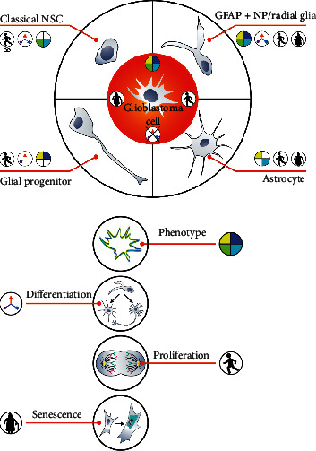
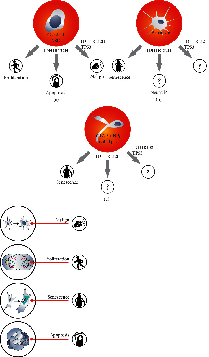
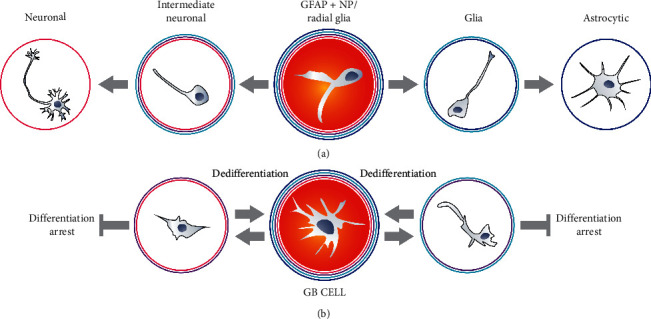
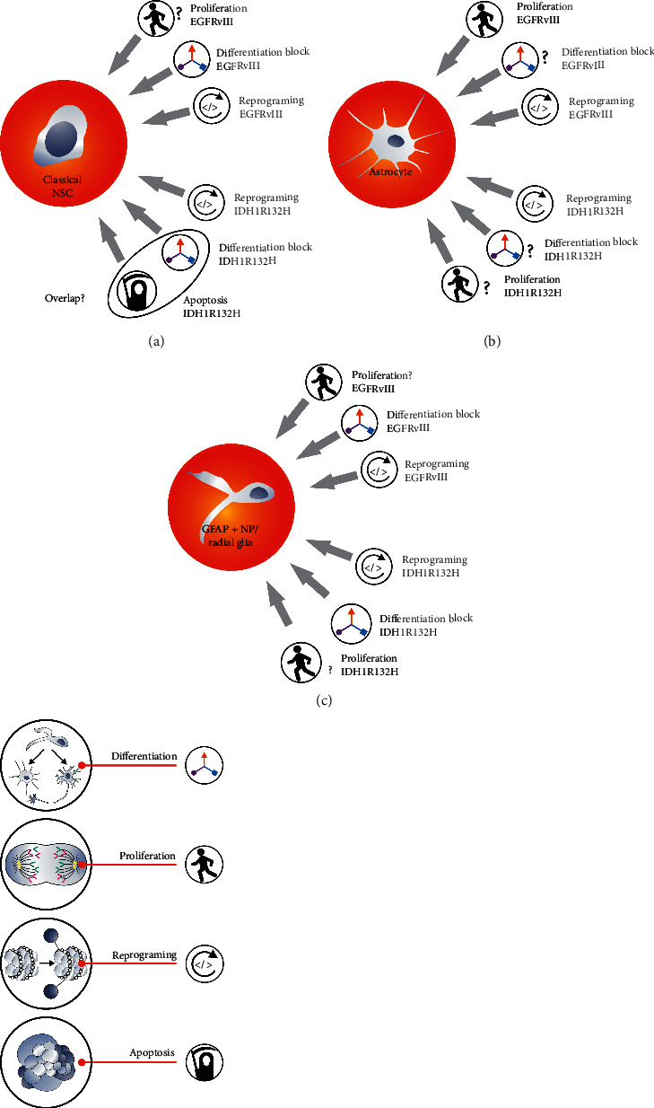
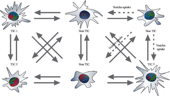
Cellular origin of glioblastoma (GB) is constantly discussed and remains a controversial subject. Unfortunately, neurobiologists are not consistent in defining neural stem cells (NSC) complicating this issue even further. Nevertheless, some suggestions referring to GB origin can be proposed based on comparing GB to central nervous system (CNS) cells. Firstly, GB cells show in vitro differentiation pattern similar to GFAP positive neural cells, rather than classical (GFAP negative) NSC. GB cells in primary cultures become senescent in vitro, similar to GFAP positive neural progenitors, whereas classical NSC proliferate in vitro infinitely. Classical NSC apoptosis triggered by introduction of IDH1R132H undermines hypothesis stating that IDH-mutant (secondary) GB origins from these NSC. Analysis of biological role of typical IDH-wildtype (primary) GB oncogene such as EGFRvIII also favors GFAP positive cells rather than classical NSC as source of GB. Single-cell NGS and single-cell transcriptomics also suggest that GFAP positive cells are GB origin. Considering the above-mentioned and other discussed in articles data, we suggest that GFAP positive cells (astrocytes, radial glia, or GFAP positive neural progenitors) are more likely to be source of GB than classical GFAP negative NSC, and further in vitro assays should be focused on these cells. It is highly possible that several populations of tumor initiating cells (TIC) exist within GB, adjusting their phenotype and even genotype to various environmental conditions including applied therapy and periodically going through different TIC states as well as non-TIC state. This adjustment is driven by changes in number and types of amplicons. The existence of various populations of TIC would enable creating neoplastic foci in different environments and increase tumor aggressiveness.


Conflict of interest statement

The authors have declared that there are no conflicts of interest regarding the publication of this article.

Figures

Figure 1
Figure 1
Similarities and differences between glioblastoma cells versus astrocytes, glial progenitors, GFAP + NP (radial glia), and classical NSC. GB cells resemble GFAP + NP the most in terms of phenotype and susceptibility to senescence. Classical NSC and astrocytes do not show the expression of GFAP and SOX2, respectively. Although GFAP + NP radial glia and GB show similar differentiation features, GB differentiation is blocked. Astrocytes and glial progenitors differentiate in the same way as GB cells. Classical NSC, unlike GB cells, proliferate in vitro far beyond the limit. GFAP + NP and GB cells quickly become senescent in vitro.
Figure 2
Figure 2
Explaining the origin of secondary glioblastoma requires IDH1R132H analysis; however, this oncogene influence on cells other than NSC was not examined profoundly.
Figure 3
Figure 3
GFAP + NP cells differentiate similarly to glioblastoma cells. However, GB cell differentiation appears to be blocked at the early stages. GFAP + NP radial glial cells also differentiate in a very distinctive way due to the presence of GFAP. The loss of GFAP is essential for NP to differentiate into neurons. GB cells exhibit similar differentiation characteristics but appear to be blocked at the early stages. GB cells, in contrast to NSC or GFAP + NP, can differentiate and dedifferentiate.
Figure 4
Figure 4
Actions of two selected and mutually exclusive oncogenes EGFRvIII and IDH1R132H suggest origin of primary and secondary glioblastoma, respectively.
Figure 5
Figure 5
Phenotypic heterogeneity of glioblastoma may result from genotypic heterogeneity associated with a different number of extrachromosomal amplicons and their different composition. Various types of tumor initiating cells can convert/transit from one type to another or to non-TIC cells, whereas non-TIC cells can convert/transit to TIC due to the changes in types of amplicons as well as their numbers and epigenetic changes. The presence of different TIC increases tumor aggressiveness, since neoplastic cells are able to invade different environments and survive many environmental changes including applied therapy. Amplicons can be transported in extracellular vesicles. Separated amplicons contain genes such as EGFR and EGFRvIII, MDM2, PDGFR, or c-MYC.

References

    1. Louis D. N., Perry A., Reifenberger G., et al. The 2016 world health organization classification of tumors of the central nervous system: a summary. Acta Neuropathologica. 2016;131(6):803–820. doi: 10.1007/s00401-016-1545-1. - DOI - PubMed
    1. Jiang Y., Marinescu V. D., Xie Y., et al. Glioblastoma cell malignancy and drug sensitivity are affected by the cell of origin. Cell Reports. 2017;18(4):977–990. doi: 10.1016/j.celrep.2017.01.003. - DOI - PubMed
    1. Bhaduri A., Di Lullo E., Jung D., et al. Outer radial glia-like cancer stem cells contribute to heterogeneity of glioblastoma. Cell Stem Cell. 2020;26(1):48–63. doi: 10.1016/j.stem.2019.11.015. - DOI - PMC - PubMed
    1. Pollard S., Conti L. Investigating radial glia in vitro. Progress in Neurobiology. 2007;83(1):53–67. doi: 10.1016/j.pneurobio.2007.02.008. - DOI - PubMed
    1. Kriegstein A., Noctor S., Martínez-Cerdeño V. Patterns of neural stem and progenitor cell division may underlie evolutionary cortical expansion. Nature Reviews Neuroscience. 2006;7(11):883–890. doi: 10.1038/nrn2008. - DOI - PubMed

LinkOut - more resources