. 2020 Oct;20(4):6.
doi: 10.3892/ol.2020.11867. Epub 2020 Jul 15.

STAT3-PTTG11 abrogation inhibits proliferation and induces apoptosis in malignant glioma cells

Affiliations

STAT3-PTTG11 abrogation inhibits proliferation and induces apoptosis in malignant glioma cells

Lishan Cui et al. Oncol Lett. 2020 Oct.

Abstract

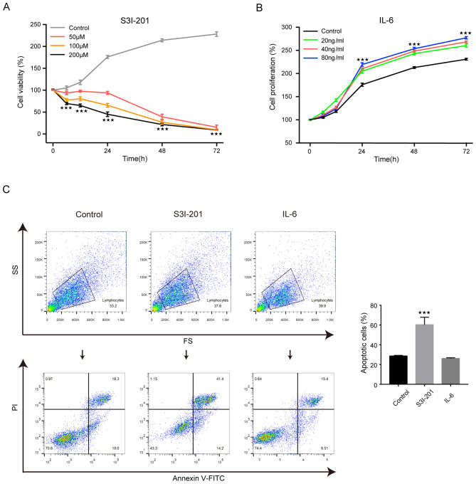
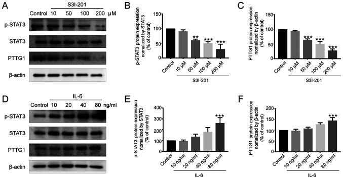
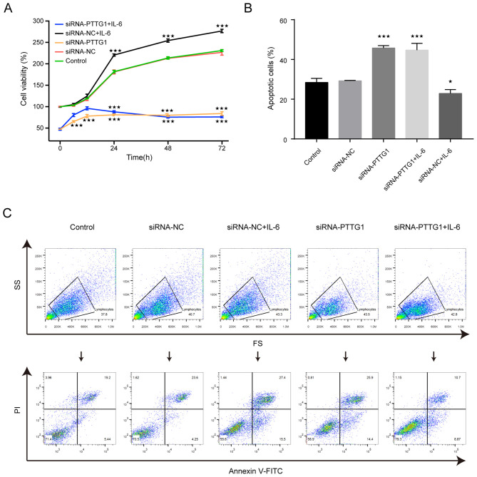
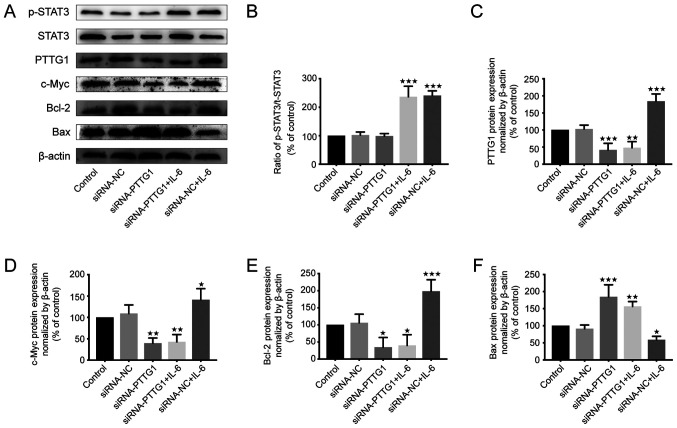
Pituitary tumor transforming gene 1 (PTTG11) is abundantly expressed in glioma. Our previous study demonstrated that the downregulation of PTTG11 gene expression significantly inhibited the proliferation, migration and invasion ability, and increased the apoptosis of SHG44 glioma cells. However, the molecular mechanisms that regulate PTTG11 and its actions remain elusive. In the present study, CCK-8 and flow cytometry assays were used to assess the proliferation/viability and apoptosis, respectively, of the human glioma U251 cell line. STAT3-PTTG1 signals were further evaluated by western blotting. The findings of the present study revealed that STAT3 induced PTTG11 expression, which subsequently induced downstream c-Myc and Bcl-2 expression while inhibiting Bax expression, thereby promoting cell viability and inhibiting apoptosis. PTTG11 suppression via siRNA inhibited the viability and increased the apoptosis of glioma cells induced by the STAT3 activator S3I-201. c-Myc and Bcl-2 expression was suppressed by PTTG11 inhibition. The findings of the present study suggest that the STAT3-PTTG11 signaling pathway may play an important role in glioma progression by regulating cell proliferation and apoptosis.

Keywords: STAT3; apoptosis; malignant glioma; pituitary tumor transforming gene 1; proliferation.


Figures

Figure 1.
Figure 1.
Effects of S3I-201 and IL-6 on proliferation and apoptosis of U251 cells. (A) Viability of U251 cells in the S3I-201 group was significantly decreased. (B) The viability of U251 cells in the IL-6 group was significantly increased. (C) S3I-201 could promote the apoptosis of U251 cells while IL-6 inhibited apoptosis. The experiment was repeated three times. ***P<0.001 compared with the control group (set to 100%). IL, interleukin; SS, side scatter; FS, forward scatter; PI, propidium iodide.
Figure 2.
Figure 2.
Effect of different doses of S3I-201 and IL-6 on the protein expression of p-STAT3 and PTTG11 proteins in U251 cells. (A) Western blotting results of p-STAT3, STAT3 and PTTG11 proteins in U251 cells treated with S3I-201. Expression levels of the (B) p-STAT3 and (C) PTTG11 proteins were both decreased after S3I-201 treatment. (D) Western blotting results of p-STAT3, STAT3 and PTTG11 proteins in U251 cells treated with IL-6. Expression levels of (E) p-STAT3 and (F) PTTG11 proteins were both increased after IL-6 treatment. Quantified results were normalized to β-actin expression. Values were expressed as percentages compared to control group (set to 100%). The experiment was repeated three times. **P<0.01, ***P<0.001 compared with the control group (set to 100%). IL, interleukin; p, phosphorylated; PTTG11, pituitary tumor transforming gene 1.
Figure 3.
Figure 3.
Effect of siRNA-PTTG11 on the viability and apoptosis of U251 cells. The control group was untreated, and negative control group cells were treated with NC siRNA. (A) Viability of U251 cells in each group after cell transfection. The viability of the U 251 cells decreased after the cells were transfected with siRNA-PTTG11, and there was no statistically significant difference between the siRNA-PTTG11 and siRNA-PTTG11+IL-6 (80 ng/ml) groups after 24 h, while the viability of the siRNA-NC+IL-6 group was significantly increased from 24 to 72 h. (B and C) Apoptosis rate of U251 cells in each group after transfection. Apoptosis was significantly increased in the siRNA-PTTG11 and siRNA-PTTG11+IL-6 groups, but decreased in the siRNA-NC+IL-6 group compared with siRNA-NC group. *P<0.05 and ***P<0.001 compared with the control group (set to 100%). IL, interleukin; SS, side scatter; FS, forward scatter; PI, propidium iodide; PTTG11, pituitary tumor transforming gene 1; NC, negative control; si, small interfering.
Figure 4.
Figure 4.
IL-6 affects apoptosis and proliferation by altering the expression of related proteins in U251 cells. The control group was untreated, and negative control group cells were treated with NC siRNA. (A) Expression of proliferation and apoptosis-related proteins in U251 cells of each group as shown by western blotting. The expression levels of p-STAT3, PTTG11, c-Myc, Bcl-2 and Bax in U251 cells after the indicated treatment. (B) Whether PTTG11 was silenced or not, IL-6 could upregulate the expression of p-STAT3. (C) IL-6 at dosage of 80 ng/ml could upregulate the expression of PTTG11. This effect was not obvious when PTTG11 was silenced. (D) c-Myc and (E) Bcl-2 were decreased in the siRNA-PTTG11 and siRNA-PTTG11+IL-6 groups, but enhanced in the siRNA-NC+IL-6 group compared with siRNA-NC group. (F) Bax levels in the siRNA-PTTG11 and siRNA-PTTG11+IL-6 groups were notably higher but were at their lowest in the siRNA-NC+IL-6 group compared with siRNA-NC group. *P<0.05, **P<0.01 and ***P<0.001 compared with the control group (set to 100%). IL, interleukin; PTTG11, pituitary tumor transforming gene 1; NC, negative control; si, small interfering.

Similar articles

Cited by

References

    1. Louis DN, Perry A, Reifenberger G, von Deimling A, Branger DF, Cavenee WK, Ohgaki H, Wiestler OD, Kleihues P, Ellison DW. The 2016 world health organization classification of tumors of the central nervous system: A summary. Acta Neuropathol. 2016;131:803–820. doi: 10.1007/s00401-016-1545-1. - DOI - PubMed
    1. Hawkins-Daarud A, Rockne RC, Anderson AR, Swanson KR. Modeling tumor-associated edema in gliomas during anti-angiogenic therapy and its impact on imageable tumor. Front Oncol. 2013;3:66. doi: 10.3389/fonc.2013.00066. - DOI - PMC - PubMed
    1. Lin L, Cai J, Jiang C. Recent advances in targeted therapy for glioma. Curr Med Chem. 2017;24:1365–1381. doi: 10.2174/0929867323666161223150242. - DOI - PubMed
    1. Omuro A, DeAngelis LM. Glioblastoma and other malignant gliomas: A clinical review. JAMA. 2013;310:1842–1850. doi: 10.1001/jama.2013.280319. - DOI - PubMed
    1. Navarro L, Gil-Benso R, Megías J, Muñoz-Hidalgo L, San- Miguel T, Callaghan RC, González-Darder JM, López-Ginés C, Cerdá-Nicolás MJ. Alteration of major vault protein in human glioblastoma and its relation with EGFR and PTEN status. Neuroscience. 2015;297:243–251. doi: 10.1016/j.neuroscience.2015.04.005. - DOI - PubMed