. 2020 Oct;20(4):28.
doi: 10.3892/ol.2020.11889. Epub 2020 Jul 17.

STAT1 expression and HPV16 viral load predict cervical lesion progression

Affiliations

STAT1 expression and HPV16 viral load predict cervical lesion progression

Si Wu et al. Oncol Lett. 2020 Oct.

Abstract

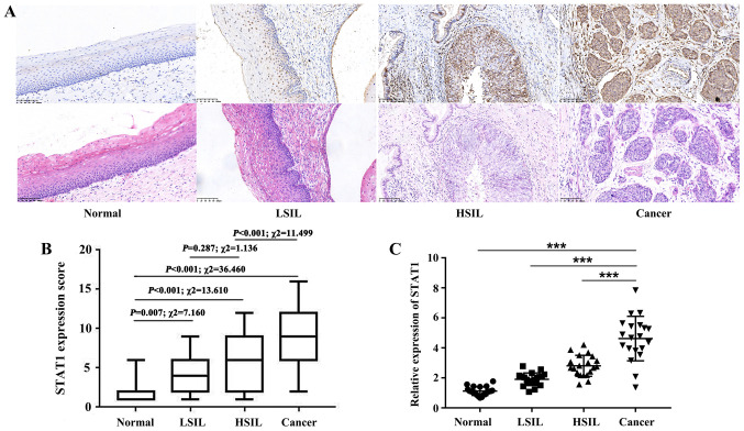
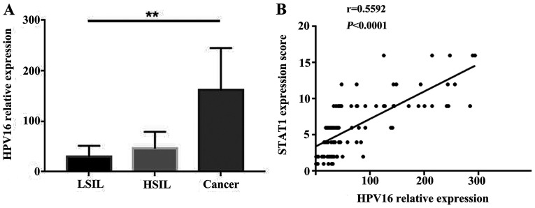
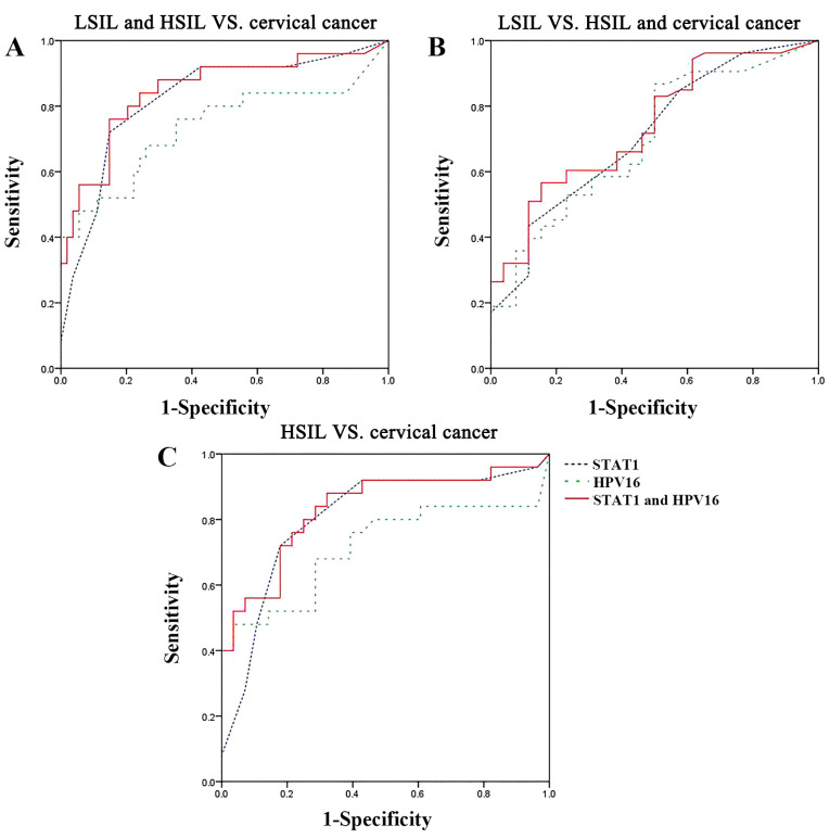
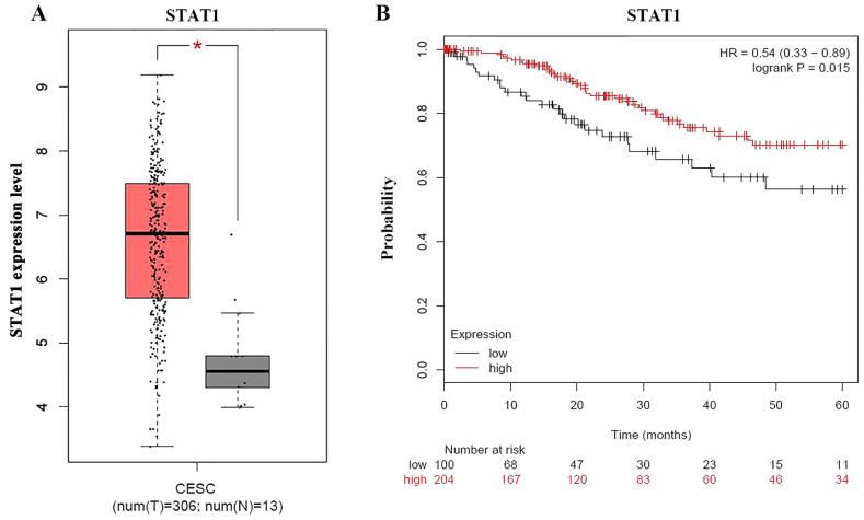
Cervical cancer is the fourth leading cause of cancer-associated mortality worldwide. However, its underlying molecular mechanisms are unclear. It is important to explore these mechanisms in order to identify novel diagnostic and prognostic biomarkers. The present study determined the association between STAT1 and human papillomavirus (HPV)16 in cervical lesions. STAT1 expression was detected by immunohistochemistry. Quantitative PCR was used to detect HPV16 viral load and STAT1 expression in cervical lesions. The potential associations among STAT1 expression, HPV16 viral load and the severity of cervical lesions in patients were analyzed using receiver operating characteristic (ROC) curves. The Cancer Genome Atlas database was used to analyze STAT1 expression and survival. High STAT1 expression was observed in 10.71 (3/28), 41.18 (14/34), 53.06 (26/49) and 90.00% (27/30) of normal tissue, low-grade squamous intraepithelial lesion (LSIL), high-grade squamous intraepithelial lesion (HSIL) and cervical squamous cell carcinoma samples, respectively. The HPV16 copy number gradually increased with the progression of cervical lesions, with the highest copy number observed in cervical cancer samples. In addition, STAT1 expression was positively correlated with HPV16 viral load. Furthermore, ROC curve analysis demonstrated that the combination of STAT1 expression and HPV16 viral load was able to differentiate between LSIL/HSIL and cervical cancer samples. Bioinformatics analysis revealed that STAT1 expression was associated with improved survival in cervical cancer. Additionally, STAT1 expression was positively associated with the progression of cervical lesions, and HPV16 viral load may affect STAT1 expression. Overall, these findings indicate that STAT1 may be an indicator of the status of cervical lesions.

Keywords: STAT1; bioinformatics; cervical lesion/cervical cancer; human papillomavirus strain 16; viral load.


Figures

Figure 1.
Figure 1.
STAT1 expression in cervical tissues. (A) Immunohistochemical staining of STAT1 protein expression in normal cervical squamous epithelium, LSIL, HSIL and cervical squamous cell carcinoma. Magnification, ×200. (B) Box plot of the immunohistochemical staining scores of STAT1 in normal tissues, LSIL, HSIL and cancer. Statistical analysis was performed using the χ2 test. (C) Total RNA was isolated to analyze STAT1 mRNA expression (20 cases per group). Data are presented as the mean ± SD. ***P≤0.001. HSIL, high-grade squamous intraepithelial lesion; LSIL, low-grade squamous intraepithelial lesion.
Figure 2.
Figure 2.
HPV16 viral load in cervical lesions. (A) HPV16 L1 reverse transcription-quantitative PCR using GAPDH as an internal control. HPV16 viral load was assessed using the 2−ΔΔCT method. One-way ANOVA followed by Dunnett's post hoc test was used for statistical analysis. (B) Correlation between HPV16 DNA load and STAT1 expression in different cervical lesions. Spearman's correlation analysis was performed. **P<0.01. HPV, human papillomavirus; HSIL, high-grade squamous intraepithelial lesion; LSIL, low-grade squamous intraepithelial lesion.
Figure 3.
Figure 3.
Receiver operating characteristic curves for the distinction between tissue samples using STAT1 expression and HPV16 viral load individually or in combination. (A) LSIL and HSIL vs. cervical cancer. (B) LSIL vs. HSIL and cervical cancer. (C) HSIL vs. cervical cancer. HPV, human papillomavirus; HSIL, high-grade squamous intraepithelial lesion; LSIL, low-grade squamous intraepithelial lesion.
Figure 4.
Figure 4.
Bioinformatics analysis of STAT1 expression in cervical tissues. (A) Box plot of STAT1 expression in CESC tissues and non-cancerous tissues. (B) Survival curves of patients with cervical cancer grouped by STAT1 expression. *P<0.05. CESC, cervical squamous cell carcinoma; HR, hazard ratio.

Similar articles

Cited by

References

    1. Cohen PA, Jhingran A, Oaknin A, Denny L. Cervical cancer. Lancet. 2019;393:169–182. doi: 10.1016/S0140-6736(18)32470-X. - DOI - PubMed
    1. Crosbie EJ, Einstein MH, Franceschi S, Kitchener HC. Human papillomavirus and cervical cancer. Lancet. 2013;382:889–899. doi: 10.1016/S0140-6736(13)60022-7. - DOI - PubMed
    1. Small W, Jr, Bacon MA, Bajaj A, Chuang LT, Fisher BJ, Harkenrider MM, Jhingran A, Kitchener HC, Mileshkin LR, Viswanathan AN, Gaffney DK. Cervical cancer: A global health crisis. Cancer. 2017;123:2404–2412. doi: 10.1002/cncr.30667. - DOI - PubMed
    1. Nitecki R, Feltmate CM. Human papillomavirus and nonhuman papillomavirus pathways to vulvar squamous cell carcinoma: A review. Curr Probl Cancer. 2018;42:476–485. doi: 10.1016/j.currproblcancer.2018.06.008. - DOI - PubMed
    1. Latsuzbaia A, Tapp J, Nguyen T, Fischer M, Arbyn M, Weyers S, Mossong J. Analytical performance evaluation of Anyplex II HPV28 and Euroarray HPV for genotyping of cervical samples. Diagn Microbiol Infect Dis. 2016;85:318–322. doi: 10.1016/j.diagmicrobio.2016.04.011. - DOI - PubMed