

Calcium pyrophosphate deposition disease: The role of imaging in their detection and in differential diagnosis of crystal arthropathies
- PMID: 32774579
- PMCID: PMC7403887
- DOI: 10.1016/j.radcr.2020.07.012
Calcium pyrophosphate deposition disease: The role of imaging in their detection and in differential diagnosis of crystal arthropathies
Abstract
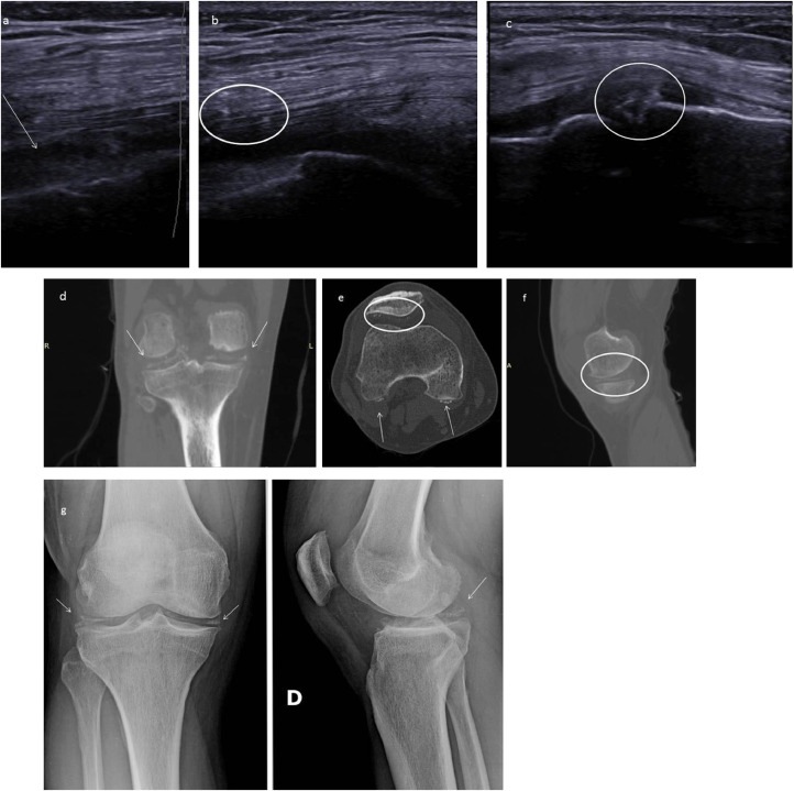
Calcium pyrophosphate deposition disease is characterized by the deposition of pyrophosphate crystals in various joint structures. Calcium pyrophosphate deposition disease can be linked to underlying metabolic disorders such as hemochromatosis, hyperparathyroidism, hypophosphatemia, hypomagnesaemia, and hypothyroidism, all of which increase the risk of calcium pyrophosphate deposition. We present the case of a 55-year-old male who underwent diagnostic examination for the onset of recurrent joint pain in the right knee.
Keywords: CPPD; CT; Chondrocalcinosis; Imaging arthropathy; Pseudogout; US.
© 2020 The Authors. Published by Elsevier Inc. on behalf of University of Washington.
Figures
Similar articles
-
Calcium Pyrophosphate Dihydrate Deposition Disease in Young Patients: Two Case Reports.Arch Rheumatol. 2016 Dec 8;32(1):80-83. doi: 10.5606/ArchRheumatol.2017.6015. eCollection 2017 Mar. Arch Rheumatol. 2016. PMID: 30375521 Free PMC article.
-
[Calcium pyrophosphate dihydrate crystal induced arthropathy].Rev Med Suisse. 2007 Mar 21;3(103):740-2, 744, 746. Rev Med Suisse. 2007. PMID: 17458152 Review. French.
-
Calcium pyrophosphate dihydrate crystal deposition disease.Semin Musculoskelet Radiol. 2003 Sep;7(3):175-85. doi: 10.1055/s-2003-43228. Semin Musculoskelet Radiol. 2003. PMID: 14593559 Review.
-
[Diagnosis and treatment of calcium pyrophosphate crystal-induced arthropathy].Z Rheumatol. 2007 Nov;66(7):573-4, 576-8. doi: 10.1007/s00393-007-0221-1. Z Rheumatol. 2007. PMID: 17932681 German.
-
[Chondrocalcinosis due to calcium pyrophosphate deposition (CPPD). From incidental radiographic findings to CPPD crystal arthritis].Z Rheumatol. 2014 May;73(4):349-57; quiz 358-9. doi: 10.1007/s00393-014-1364-5. Z Rheumatol. 2014. PMID: 24811359 Review. German.
Cited by
-
Reducing diagnostic delay in hypophosphatasia: a case series of 14 patients presenting to general rheumatology.Osteoporos Int. 2023 Sep;34(9):1647-1652. doi: 10.1007/s00198-023-06749-z. Epub 2023 Apr 28. Osteoporos Int. 2023. PMID: 37118032 Free PMC article.
-
Degenerative lesion of the extensor tendon V finger: Ultrasound imaging in diagnosis and therapeutic possibilities.Eur J Radiol Open. 2021 May 3;8:100348. doi: 10.1016/j.ejro.2021.100348. eCollection 2021. Eur J Radiol Open. 2021. PMID: 34012998 Free PMC article.
References
-
- Lindenmeyer C., Sobel A., Nazarian L., Mandel S., Raikin S. A case study of pseudo-neuropathic pseudogout. Med Forum. 2013;14(26)
-
- Yochum T., Rowe L. Williams & Wilkins; Baltimore: 1996. Essentials of skeletal radiology.
-
- Abhishek A. Calcium pyrophosphate deposition. Br J Hosp Med. 2014;75(4):C61–C64. - PubMed
Publication types
LinkOut - more resources
Full Text Sources
