. 2020 Jul 28;11(30):2873-2888.
doi: 10.18632/oncotarget.27556.

Th1 cytokines in conjunction with pharmacological Akt inhibition potentiate apoptosis of breast cancer cells in vitro and suppress tumor growth in vivo

Affiliations

Th1 cytokines in conjunction with pharmacological Akt inhibition potentiate apoptosis of breast cancer cells in vitro and suppress tumor growth in vivo

Loral Showalter et al. Oncotarget. .

Abstract

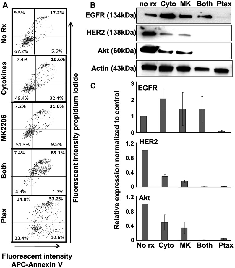
Targeted drug approaches have been a major focus for developing new anticancer therapies. Although many such agents approved in the last 20 years have improved outcomes, almost all have underperformed expectations. The full potential of such agents may yet be obtained through novel combinations. Previously, we showed that anti-estrogen drugs combined with a dendritic cell-based anti-HER-2 vaccine known to induce strong Th1-polarized immunity dramatically improved clinical response rates in patients with HER-2pos/ERpos early breast cancer. Here, we show that the small molecule Akt antagonist MK-2206, when combined with the Th1 cytokines IFN-gamma and TNF-alpha, maximize indicators of apoptotic cell death in a panel of phenotypically-diverse human breast cancer lines. These findings were mirrored by other, structurally-unrelated Akt-targeting drugs that work through different mechanisms. Interestingly, we found that MK-2206, as well as the other Akt antagonist drugs, also had a tendency to suppress Th1 cytokine expression in stimulated human and murine lymphocytes, potentially complicating their use in conjunction with active immunotherapy. After verifying that MK-2206 plus IFN-gamma could show similar combined effects against breast cancer lines, even in the absence of TNF-alpha, we tested in a rodent HER-2pos breast cancer model either a HER-2-based DC vaccine, or recombinant IFN-gamma with or without MK-2206 administration. We found that for MK-2206, co-administration of recombinant IFN-gamma outperformed co-administration of DC vaccination for slowing tumor growth kinetics. These findings suggest a combined therapy approach for Akt-targeting drugs that incorporates recombinant Interferon-gamma and is potentially translatable to humans.

Keywords: Akt kinase; breast cancer; immunotherapy.


Conflict of interest statement

CONFLICTS OF INTEREST None.

Figures

Figure 1
Figure 1. MK-2206 plus Th1 cytokines potentiate suppression of metabolic activity for human breast cancer cells.
SKBR-3, MDA-MB-468, MDA-MB-453 and HCC-1419 cells were seeded at 5 × 10³ per well in 96-well cluster plate and cultured overnight. The cells were then exposed to Th1 cytokines (IFN-γ and TNF-α at 10 ng/ml each), MK-2206 (10 μM), both treatments, or left untreated, and incubated for an additional 72 h. Then, 20 μl of resazurin sodium salt solution (1.4 mg/ml) was added per well, and cells incubated until color change occurred. Optical density of culture supernatants was determined at 630 nm. Experiments were repeated at least three times for each cell line. Error bars depict the SEM.
Figure 2
Figure 2. MK-2206 plus Th1 cytokines enhances cell death for breast cancer lines.
SKBR-3, MDA-MB-468, MDA-MB-453 and HCC-1419 cells were seeded at 1 × 10⁵ per well in 12-well cluster plates and cultured overnight. The following day cells were then exposed to Th1 cytokines (IFN-γ and TNF-α at 10 ng/ml each), MK-2206 (10 μM), both treatments, or left untreated. After 72 h further incubation, cells were then harvested, washed and stained with Trypan Blue dye, then analyzed by flow cytometry for dye uptake. (A) Histogram analysis of a representative experiment. A region was defined to enumerate the percentage of dead (high-fluorescence, Trypan Blue-stained) cells. (B) Statistical analysis of composite data from at least 3 separate experiments per cell line using mean channel fluorescence. Error bars denote SEM.
Figure 3
Figure 3. MK-2206 plus Th1 cytokines potentiate mitochondrial depolarization in human breast cancer lines.
SKBR-3, MDA-MB-468, MDA-MB-453 and HCC-1419 cells were cultured at 1 × 10⁵ per well in 12-well cluster plates overnight. The next day cells were then exposed to Th1 cytokines (IFN-γ and TNF-α at 10 ng/ml each), MK-2206 (10 μM), both treatments, or left untreated. After 72 h further incubation, TMRE dye (final concentration 100 nM) was added to the cells which were incubated for an additional 20 minutes, after which cells were harvested, washed, resuspended in PBS and subjected to flow cytometry analysis. (A) Histogram analysis depicting a representative experiment. A region was defined to enumerate the percentage of low-staining (mitochondria-depolarized) cells. (B) Statistical analysis of composite data from at least 3 separate experiments using mean channel fluorescence. Error bars denote SEM.
Figure 4
Figure 4. Combined MK-2206 and Th1 cytokines maximize markers of cellular apoptosis for human breast cancer cell lines.
SKBR-3, MDA-MB-468, MDA-MB-453 and HCC-1419 cells were cultured at 1 × 10⁵ per well in 12-well cluster plates overnight. Cells were then exposed to Th1 cytokines (IFN-γ and TNF-α at 10ng/ml each), MK-2206 (10 μM), both treatments, or left untreated. After an additional 72 h incubation, cells were harvested, washed and resuspended in binding buffer, and then stained with propidium iodide (PI) and APC-labeled Annexin V. Stained cells were subjected to flow cytometry analysis. Quadrants were defined to differentiate live, double-negative cells (lower left quadrant) from apoptotic, double-positive cells (upper right quadrant), and establish percentages of cells within these categories. Data shown is one representative experiment from at least 3 individual trials for each cell line.
Figure 5
Figure 5. Th1 cytokines, MK-2206, and paclitaxel induce apoptosis in human breast cancer cells but differentially affect expression of oncodrivers.
SKBR3 cells were seeded at a density of 1 × 10⁵ per well in 12-well cluster plates, and cultured overnight. The next day cells were exposed to Th1 cytokines (IFN-γ and TNF-α at 10 ng/ml each), MK-2206 (10 μM), both treatments, paclitaxel (300 nM) or left untreated. After 72 h further incubation, cells were harvested and (A) stained with propidium iodide and APC-labeled Annexin V. Stained cells were subjected to flow cytometry and data evaluated by quadrant analysis, with double-staining cells (upper right quadrant) defined as apoptotic. (B) Western blot analysis of cell extracts for expression of EGFR, HER2, Akt kinase and actin (loading control). (C) Densitometry analysis of western blots (composite data from 3 separate experiments) normalized to actin expression. Error bars indicate SEM.
Figure 6
Figure 6. MK-2206 suppresses IFN-γ responses in human PBMCs and murine splenocytes.
(A) Cryopreserved human PBMCs from healthy donors were thawed and plated at a density of 3 × 10⁵ per well in human IFN-γ 96-well Elispot plates. Cells were then stimulated with either a mixture of peptides based on the sequence of common viral antigens (upper panels) or tetanus toxoid (lower panels) in the presence or absence of MK2206 (10 μM). Elispot plates were incubated overnight, then developed and IFN-γ spot forming cells enumerated. Data expressed as the percentage of the positive control (left panels), with photographs of representative, developed wells also shown (right panels). (B) Freshly prepared mouse splenocytes were cultured in IFN-γ elispot plates at 3 × 10⁵ per well and then exposed to mitogen (concanavilin A) in the presence or absence of MK2206 (10 μM). Cells were incubated overnight and the next morning plates were developed and IFN-γ spot forming cells enumerated. Left panel represent total IFN-γ spots obtained with various treatments, with error bars denoting SEM from triplicate wells. Right panels show photographs of representative developed wells.
Figure 7
Figure 7. Other Akt antagonists display similar apoptosis-inducing and T-cell inhibitory properties as MK-2206.
(A) MDA-MB-453 and MDA-MB-468 cells were cultured overnight in 96-well plates. The next day they were treated with perifosine (20 μM), GSK-690693 (10 μM), or GDC-068 (40 μM) in the presence or absence of Th1 cytokines (IFN-γ and TNFα at 10ng/ml each). After 72 hours of treatment, 20 μl resazurin sodium salt solution (1.4 mg/ml) was added to each well and the cells were incubated until color change occurred. Optical densities were read spectrophotometrically at 630 nM. Shown are composite data from at least three trials per cell line expressed as mean optical density +/– SEM. (B) An IFN-γ ELISPOT was performed according to manufacturers’ instructions using PBMCs from 5 different donors (left panel). The PBMCs were cultured with viral recall peptides overnight in the presence of MK2206 (10 μM), Perifosine (20 μM), GSK690693 (10 μM), or left untreated for control. The next day the number of spot forming cells were counted and the data expressed as the percent of the control. IFN-γ ELISA analysis of 48-hour culture supernatants from allogeneic MLRs where activated dendritic cells (DC) and lymphocyte-rich elutriation fractions were co-cultured at 1:40 stimulator: responder ratios in the presence or absence of drug. Data displayed represents the percent maximum of mean IFN-γ production from seven unique allogeneic DC: lymphocyte pairings.
Figure 8
Figure 8. MK-2206 in conjunction with immunotherapy slows progression of rodent HER-2pos tumors.
(A) Erbb2pos (rat homolog of HER-2) TUBO cells were cultured overnight in 96-well culture plates. The next day they were exposed to increasing concentrations of MK2206 in the presence (solid circles) or absence (dotted triangles) of IFN-γ (50 ng/ml) and incubated a further 72 h, after which 20 μl of resazurin sodium salt solution (1.4 mg/ml) was added to each well, and upon color change, optical density of culture supernatants assessed at 630 nm (left panel). Cells similarly treated were also assessed by Trypan Blue staining, and dye uptake evaluated by flow cytometry (right panel). Error bars indicate SEM. (B) 5 × 10⁵ TUBO cells were implanted into the fat pad of one of the right mammary glands of female Balb/c mice. When tumors became palpable (seven days), mice were provided with six treatment regimens (5 mice per group) including no treatment (upper and lower panels), MK2206 (upper and lower panels), rat HER-2 peptide-pulsed dendritic cells (upper panels), peptide-pulsed DCs plus MK2206 (upper panels), IFN-γ (lower panels) and IFN-γ plus MK2206 (lower panel). MK2206 was supplied in two 5-day cycles separated by a 2 day rest period. IFN-γ was supplied on the same schedule as MK-2206. DCs were administered twice weekly during cycles of MK2206 treatment. Tumor growth was monitored 2–3 times per week. Growth curves are displayed in left panels, and statistical analysis at the terminus of treatment displayed in right panels. Error bars indicate SEM.

Similar articles

Cited by

References

    1. Oiseth SJM, Aziz S. Cancer Immunotherapy: a brief review of the history, possibilities, and challenges ahead. J Cancer Metastasis Treat. 2017; 3:250–61. 10.20517/2394-4722.2017.41. - DOI
    1. Czerniecki B, Koski G, Koldovsky U, Xu S, Cohen P, Mick R, Nisenbaum H, Pasha T, Xu M, Fox K, Weinstein S, Orel S, Vonderheide R, et al.. Targeting HER-2/neu in early breast cancer development using dendritic cells with staged interleukin-12 burst secretion. Cancer Res. 2007; 67:1842–52. 10.1158/0008-5472.can-06-4038. - DOI - PubMed
    1. Sharma A, Koldovsky U, Xu S, Mick R, Roses R, Fitzpatrick E, Weinstein S, Nisenbaum H, Levine BL, Fox K, Zhang P, Koski G, Czerniecki BJ. HER-2 pulsed dendritic cell vaccine can eliminate HER-2 expression and impact ductal carcinoma in situ . Cancer. 2012; 118:4354–62. 10.1002/cncr.26734. - DOI - PMC - PubMed
    1. Koski GK. A novel dendritic cell-based immunization approach for the induction of durable Th1-polarized anti-HER-2/neu responses in women with early breast cancer. Journal of Immunotherapy. 2012; 35:54–65. 10.1097/CJI.0b013e318235f512. - DOI - PMC - PubMed
    1. Braumüller HH. T-helper-1-cell cytokines drive cancer into senescence. Nature. 2013; 494:361–5. 10.1038/nature11824. - DOI - PubMed