. 2020 Aug 3:11:15.
doi: 10.1186/s13227-020-00162-x. eCollection 2020.

Expression of smooth muscle-like effectors and core cardiomyocyte regulators in the contractile papillae of Ciona

Affiliations

Expression of smooth muscle-like effectors and core cardiomyocyte regulators in the contractile papillae of Ciona

Christopher J Johnson et al. Evodevo. .

Abstract

Background: The evolution of vertebrate smooth muscles is obscured by lack of identifiable smooth muscle-like cells in tunicates, the invertebrates most closely related to vertebrates. A recent evolutionary model was proposed in which smooth muscles arose before the last bilaterian common ancestor, and were later diversified, secondarily lost or modified in the branches leading to extant animal taxa. However, there is currently no data from tunicates to support this scenario.

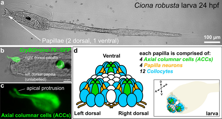
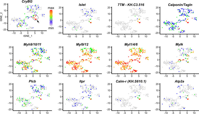
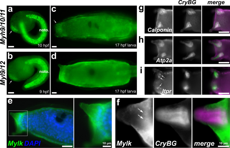
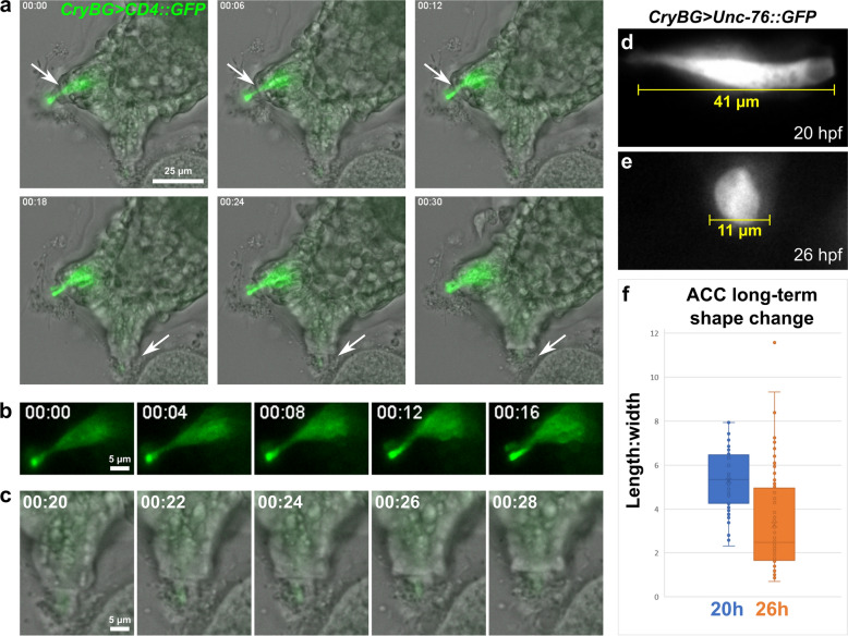
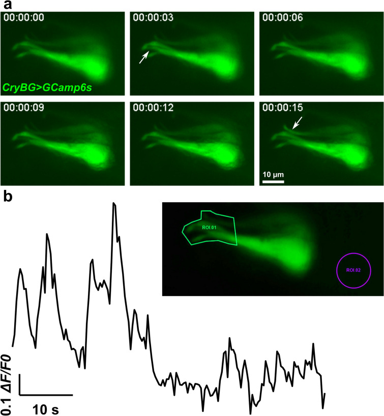
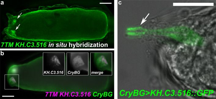
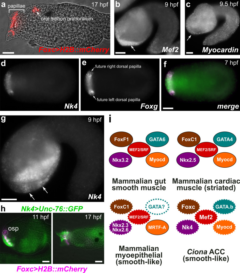
Methods and results: Here, we show that the axial columnar cells, a unique cell type in the adhesive larval papillae of the tunicate Ciona, are enriched for orthologs of vertebrate smooth/non-muscle-specific effectors of contractility, in addition to developing from progenitors that express conserved cardiomyocyte regulatory factors. We show that these cells contract during the retraction of the Ciona papillae during larval settlement and metamorphosis.

Conclusions: We propose that the axial columnar cells of Ciona are a myoepithelial cell type required for transducing external stimuli into mechanical forces that aid in the attachment of the motile larva to its final substrate. Furthermore, they share developmental and functional features with vertebrate myoepithelial cells, vascular smooth muscle cells, and cardiomyocytes. We discuss these findings in the context of the proposed models of vertebrate smooth muscle and cardiomyocyte evolution.


Conflict of interest statement

Competing interestsThe authors declare that they have no competing interests.

Figures

Fig. 1
Fig. 1
The papillae of Ciona larvae. a A Ciona robusta (intestinalis Type A) larva, showing the 3 papillae. b Larva electroporated with the reporter plasmid CryBG > Unc-76::GFP (Cirobu.REG.KhS605. 16,789–17,833, Shimeld et al., which labels axial columnar cells (ACCs) and unrelated otolith cell. This larva is a transgenic mosaic, so the left dorsal papilla is unlabeled. The ventral papilla is also unlabeled but out of focus. c Magnified view of labeled papilla in b, without brightfield overlay, showing ACCs labeled by CryBG > Unc-76::GFP expression. Each has a characteristic apical, finger-like protrusion that extends through a fenestration of the larval cellulose tunic around the apical tip of the papilla. d Cartoon diagram of the major cell types of the three papillae and their approximate arrangement in the larva. Cell types are identified by color code and described in reference [121]. Cells of unknown number and type in between the three papillae are represented in white. Sizes not to scale
Fig. 2
Fig. 2
Single-cell RNAseq differential gene expression “maps” of ACC markers. tSNE plots of re-clustered larval cells from Sharma et al. [91]. ACC-containing subcluster (arrow pointing to red- and orange-colored cells) is marked by high expression of Beta/gamma Crystallin (CryBG), and Islet, based on previous findings (see text for detail). Cells colored by relative gene expression, normalized as maximum (red) to minimum (grey) values for each gene, as indicated by color scale at top left. See also Additional file 1: Fig. S1
Fig. 3
Fig. 3
In situ hybridization for smooth muscle-like effector gene expression in ACCs. aMyh9/10/11 at 10 h post-fertilization (hpf), showing expression in papilla progenitors (arrow). Expression is also strong in the notochord (noto.). bMyl9/12 at 9 hpf, showing expression in papilla progenitors (arrow) and notochord. cMyh9/10/11 expression in 17 hpf larvae, showing faint but broad signal in the papilla territory (arrow). dMyl9/12 expression in 17 hpf larvae, showing almost undetectable expression above background in papillae, a bit more in notochord. eMylk expression (green) at 17 hpf, counterstained with DAPI to label DNA (blue). Inset of boxed area shown right, indicating broad expression in the papilla territory. f Two-color in situ hybridization with Mylk (green) and CryBG (magenta) probes show strong Mylk expression in cells surrounding the protuberances, but also more weakly in the ACCs, which are labeled specifically by CryBG. Sub-cellular mRNA localization of Mylk appears quite different from that of CryBG, but still appears to localize around nuclei of ACCs (arrows). Top right: Two-color hybridization of CryBG (green) and gCalponin,hAtp2a, and iItpr (all magenta) showing ACC-specific expression. Nuclear dots (arrows) indicate likely active transcription of Itpr. All scale bars = 25 µm unless otherwise annotated
Fig. 4
Fig. 4
The contractility and shape change of ACCs during papilla retraction. a Still images captured from live time-lapse video (one animal recorded, see Additional file 4: Video S1) of attached larva ~ 21 h post-fertilization (hpf). ACCs marked by CryBG > CD4::GFP expression. Right dorsal papilla (top arrows) retracts from 6 to 12 min timepoints, while unlabeled left dorsal papilla (bottom arrows) retracts from 18 to 24 min. b Same GFP-labeled papilla in a, but only GFP channel shown, revealing ACC contraction. c Same unlabeled papilla in a, showing the finger-like apical protrusion of the ACC retracting into papilla. f Representative image of an ACC labeled by CryBG > Unc-76::GFP at 20 hpf, indicating typical length at this stage prior to settlement. e representative image of labeled ACC at 26 hpf, indicating extreme rounded shape observed in many larvae at this stage, after settlement and metamorphosis begins. f Quantification of length:width ratio of ACCs sampled from 20 vs. 26 hpf larvae (n = 54 for 20 hpf, n = 84 for 26 hpf)
Fig. 5
Fig. 5
Intracellular calcium imaging in ACCs. a Still images captured from live time-lapse video (see Additional file 5: Video S2) of one larva electroporated with CryBG > GCamp6s, which drives expression of the calcium indicator protein GCamp6s. Transient increases in fluorescence intensity is seen in apical protrusions (arrows) around 3 s and 15 s in different cells. b Quantification of change in fluorescence in same time-lapse video, in the region of interest (ROI) 1 surrounding the apical protrusions of the ACCs (see inset), given as ΔF/F0 normalized to the maximal absolute value (1.0). See Additional file 6 for raw and normalized data
Fig. 6
Fig. 6
Expression of a putative orphan G protein-coupled receptor in the ACCs. a In situ hybridization showing expression of seven-transmembrane (7TM) protein-encoding gene KH.C3.516 in the ACCs (arrows). b Two-color in situ hybridization with KH.C3.516 (magenta) and CryBG (green) shows highly specific, strong expression in the ACCs (insets). c Expression of a KH.C3.516::GFP fusion driven by the CryBG promoter shows enriched localization in the apical tips of ACC protrusions (arrows), which are exposed to the environment through the tunic. All scale bars = 25 µm
Fig. 7
Fig. 7
Cardiomyocyte/smooth muscle-like core regulatory complex (CoRC) factors in the ACCs. a Larva electroporated with Foxc > H2B::mCherry (Cirobu.REG.KhL57. 96,067-98,197, red), recapitulating expression of Foxc in the cell lineage giving rise to both the oral siphon primordium and the papillae. b In situ hybridization for Mef2, showing broad, possibly maternal expression throughout the embryo at 9 hpf, but also upregulation in the papilla territory (arrow). c In situ hybridization for Myocardin, which is expressed ubiquitously at 9.5 hpf, including in papilla territory (arrow). df Two-color in situ hybridization at 7 hpf for dNk4 and eFoxg. Presumptive protuberances of the papillae (which include the ACCs) are marked by Foxg expression at this stage, surrounding a territory of Foxg-negative cells. f Merged image shows Nk4 (green) expressed broadly in both Foxg +(magenta) and Foxg-negative cells within the whole papilla territory. g In situ hybridization for Nk4 at 9 hpf, showing gradual restriction to three presumptive papilla protuberances, two of which are visible at this focal plane (arrows). h Electroporation of Nk4 (Cirobu.REG 4,056,723-4,057,773, green) and Foxc (magenta) reporter plasmids confirms expression of Nk4 throughout papilla territory but not the oral siphon primordium (osp), viewed at 11 hpf and 17 hpf. Oral siphon primordium not visible in 17 hpf image. Nk4 reporter mosaicism resulted in one unlabeled papilla. i Diagram comparing CoRCs for gut smooth muscles, cardiomyocytes, and myoepithelia to Ciona ACCs, suggesting deep homology according to the model proposed by Brunet et al. [8]. Dashed outline around solid color indicate expression data only, no functional data yet obtained. Dashed outline around white indicate no expression or functional data so far. All scale bars = 25 µm

Similar articles

Cited by

References

    1. Abitua PB, Gainous TB, Kaczmarczyk AN, Winchell CJ, Hudson C, Kamata K, Nakagawa M, Tsuda M, Kusakabe TG, Levine M. The pre-vertebrate origins of neurogenic placodes. Nature. 2015;524:462. - PMC - PubMed
    1. Abitua PB, Wagner E, Navarrete IA, Levine M. Identification of a rudimentary neural crest in a non-vertebrate chordate. Nature. 2012;492:104. - PMC - PubMed
    1. Anderson PJ, Lynch TJ, Engelhardt JF. Multipotent myoepithelial progenitor cells are born early during airway submucosal gland development. Am J Respir Cell Mol Biol. 2017;56:716–726. - PMC - PubMed
    1. Arendt D, Musser JM, Baker CVH, Bergman A, Cepko C, Erwin DH, Pavlicev M, Schlosser G, Widder S, Laubichler MD, Wagner GP. The origin and evolution of cell types. Nat Rev Genet. 2016;17:744–757. - PubMed
    1. Beh J, Shi W, Levine M, Davidson B, Christiaen L. FoxF is essential for FGF-induced migration of heart progenitor cells in the ascidian Ciona intestinalis. Development. 2007;134:3297–3305. - PubMed

LinkOut - more resources