

Reliability of foveal avascular zone measurements in eyes with retinal vein occlusion using optical coherence tomography angiography
- PMID: 32774887
- PMCID: PMC7398327
- DOI: 10.1186/s40942-020-00237-w
Reliability of foveal avascular zone measurements in eyes with retinal vein occlusion using optical coherence tomography angiography
Abstract
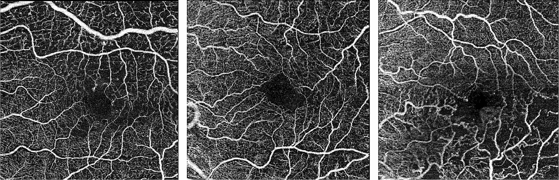
Background: To evaluate the reliability of foveal avascular zone (FAZ) area measurements using optical coherence tomography angiography (OCTA) in eyes with retinal vein occlusion (RVO).
Methods: Twenty-five OCTA exams of patients with RVO were evaluated retrospectively. Three examiners performed manual measurements of the FAZ, and interrater and intrarater reliability were obtained.
Results: The intraclass correlation coefficient (ICC) for interrater reliability for individual measurements was 0.62 (moderate) with a 95% confidence interval (CI) of 0.40 to 0.79 (p < 0.001). The ICC (95% CI) for intrarater reliability was 0.92 (0.82 to 0.96) for rater A, 0.96 (0.91 to 0.98) for B, and 0.88 (0.76 to 0.94) for C (p < 0.001). In all subanalyses including presence of edema and type of occlusion, interrater reliability was poor/moderate, and intrarater reliability was good/excellent.
Conclusion: The FAZ varies significantly among eyes with RVO, so measurements obtained using OCTA should be analyzed with caution due to the moderate level of reliability among different examiners.
Keywords: Foveal avascular zone; OCT; Retina; Retinal vein occlusion.
© The Author(s) 2020.
Conflict of interest statement
Competing interestsThe authors declare that they have no competing interests.
Figures
Similar articles
-
Evaluation of macular microvasculature and foveal avascular zone in patients with retinal vein occlusion using optical coherence tomography angiography.Int Ophthalmol. 2022 Jan;42(1):211-218. doi: 10.1007/s10792-021-02015-5. Epub 2021 Aug 22. Int Ophthalmol. 2022. PMID: 34423405
-
Comparison of foveal avascular zone between optical coherence tomography angiography and fluorescein angiography in patients with retinal vein occlusion.PLoS One. 2019 Jun 4;14(6):e0217849. doi: 10.1371/journal.pone.0217849. eCollection 2019. PLoS One. 2019. PMID: 31163058 Free PMC article.
-
CORRELATION OF MICROVASCULAR STRUCTURES ON OPTICAL COHERENCE TOMOGRAPHY ANGIOGRAPHY WITH VISUAL ACUITY IN RETINAL VEIN OCCLUSION.Retina. 2017 Sep;37(9):1700-1709. doi: 10.1097/IAE.0000000000001403. Retina. 2017. PMID: 27828907
-
[Foveal avascular zone and OCT angiography. An overview of current knowledge].Ophthalmologe. 2019 Jul;116(7):610-616. doi: 10.1007/s00347-018-0838-2. Ophthalmologe. 2019. PMID: 30569234 Review. German.
-
Optical Coherence Tomography Angiography in Eyes with Retinal Vein Occlusion.J Ophthalmic Vis Res. 2018 Jul-Sep;13(3):315-332. doi: 10.4103/jovr.jovr_264_17. J Ophthalmic Vis Res. 2018. PMID: 30090189 Free PMC article. Review.
Cited by
-
OCTA measurements in Behcet's disease across different stages of the disease activity: A systematic review and meta-analysis.PLoS One. 2025 Jul 2;20(7):e0323192. doi: 10.1371/journal.pone.0323192. eCollection 2025. PLoS One. 2025. PMID: 40601709 Free PMC article.
-
Multimodal Imaging of Microvascular Abnormalities in Retinal Vein Occlusion.J Clin Med. 2021 Jan 21;10(3):405. doi: 10.3390/jcm10030405. J Clin Med. 2021. PMID: 33494354 Free PMC article. Review.
-
Ophthalmic Biomarkers for Alzheimer's Disease: A Review.Front Aging Neurosci. 2021 Sep 10;13:720167. doi: 10.3389/fnagi.2021.720167. eCollection 2021. Front Aging Neurosci. 2021. PMID: 34566623 Free PMC article. Review.
-
Analyses of Foveal Avascular Zone in Patients with General Blunt Ocular Trauma Using Optical Coherence Tomography Angiography.Korean J Ophthalmol. 2023 Feb;37(1):62-69. doi: 10.3341/kjo.2022.0081. Epub 2023 Feb 3. Korean J Ophthalmol. 2023. PMID: 36796347 Free PMC article.
-
Eye-tracking paradigms for the assessment of mild cognitive impairment: a systematic review.Front Psychol. 2023 Jul 20;14:1197567. doi: 10.3389/fpsyg.2023.1197567. eCollection 2023. Front Psychol. 2023. PMID: 37546488 Free PMC article. Review.
References
-
- Adhi M, Filho MAB, Louzada RN, Kuehlewein L, de Carlo TE, Baumal CR, et al. Retinal capillary network and foveal avascular zone in eyes with vein occlusion and fellow eyes analyzed with optical coherence tomography angiography. Investig Opthalmol Vis Sci. 2016;57(9):OCT486. - PubMed
-
- Kang J-W, Yoo R, Jo YH, Kim HC. Correlation of microvascular structures on optical coherence tomography angiography with visual acuity in retinal vein occlusion. Retina. 2017;37(9):1700–1709. - PubMed
-
- Wons J, Pfau M, Wirth MA, Freiberg FJ, Becker MD, Michels S. Optical coherence tomography angiography of the foveal avascular zone in retinal vein occlusion. Ophthalmologica. 2016;235(4):195–202. - PubMed
-
- Novais EA, Waheed NK. Optical coherence tomography angiography of retinal vein occlusion. In: Bandello F, Souied EH, Querques G, editors. Developments in ophthalmology. S. Karger AG; 2016. p. 132–8. Accessed 18 Aug 2018. - PubMed
-
- Spaide RF, Klancnik JM, Jr, Cooney MJ. Retinal vascular layers imaged by fluorescein angiography and optical coherence tomography angiography. JAMA Ophthalmol. 2015;133(1):45–50. - PubMed
LinkOut - more resources
Full Text Sources
