Effects of Combinations of Ophthalmic Viscosurgical Devices and Suction Flow Rates on the Corneal Endothelial Cell Damage Incurred during Phacoemulsification
- PMID: 32774899
- PMCID: PMC7391086
- DOI: 10.1155/2020/2159363
Effects of Combinations of Ophthalmic Viscosurgical Devices and Suction Flow Rates on the Corneal Endothelial Cell Damage Incurred during Phacoemulsification
Abstract
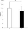
We examined the effects of different ophthalmic viscosurgical devices (OVDs) and suction flow rates during phacoemulsification on the amount of ultrasound power used and damage to the corneal endothelium. In total, 48 eyes of 24 patients who underwent phacoemulsification and intraocular lens insertion with different OVD settings in the left and right eye between February and August 2018 were examined retrospectively from medical records. Each of the following types of OVDs was used in either the right or left eye of each patient: a viscoadaptive OVD (V group) or a combination of dispersive and cohesive OVDs (soft-shell technique; S group). There was no significant difference in the lens nucleus hardness between the two groups. A 2.4 mm transconjunctival scleral incision was made, and phacoemulsification was performed by the same surgeon. The cumulative dissipated energy (CDE) and ultrasound time intraoperatively were compared between the two groups. The CDE was significantly larger in the V group (9.9 ± 4.6) than the S group (6.4 ± 3.0; p=0.006). The reduction rate of the endothelial cell density at the center of the cornea was significantly higher in the V group (4.1% ± 6.7%) than the S group (0.3% ± 4.5%; p=0.03) at 1 week postoperatively. Both groups had a good postoperative course. There was less corneal endothelial damage with the soft-shell technique combined with a normal flow setting than the viscoadaptive OVD combined with a low flow setting.
Copyright © 2020 Tomoyuki Kunishige and Hiroshi Takahashi.
Conflict of interest statement
The authors declare that they have no conflicts of interest.
Figures


References
-
- Shimmura S., Tsubota K., Oguchi Y., Fukumura D., Suematsu M., Tsuchiya M. Oxiradical–dependent photoemission induced by a phacoemulsification probe. Investigative Ophthalmology & Visual Science. 1992;33(10):2904–2907. - PubMed
LinkOut - more resources
Full Text Sources

