Review
. 2020 Jul 22:2020:3509064.
doi: 10.1155/2020/3509064. eCollection 2020.

Objective Imaging Diagnostics for Dry Eye Disease

Affiliations
Review

Objective Imaging Diagnostics for Dry Eye Disease

Sang Beom Han et al. J Ophthalmol. .

Abstract

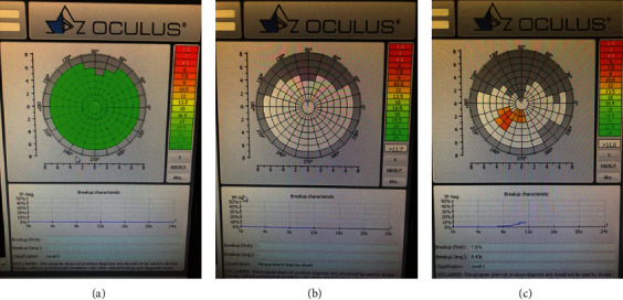
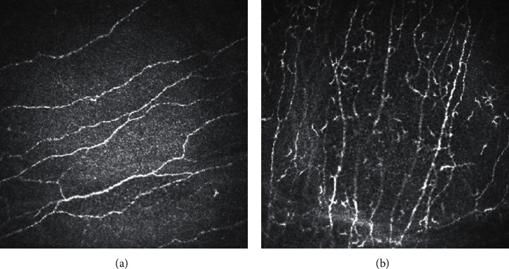
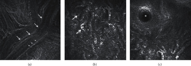
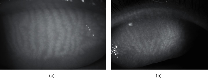
Traditional diagnostic tests for dry eye disease (DED), such as fluorescein tear film break-up time and the Schirmer test, are often associated with poor reproducibility and reliability, which make the diagnosis, follow-up, and management of the disease challenging. Advances in ocular imaging technology enables objective and reproducible measurement of changes in the ocular surface, tear film, and optical quality associated with DED. In this review, the authors will discuss the application of various imaging techniques, such as, noninvasive tear break-up time, anterior segment optical coherence tomography, in vivo confocal microscopy, meibography, interferometry, aberrometry, thermometry, and tear film imager in DED. Many studies have shown these devices to correlate with clinical symptoms and signs of DED, suggesting the potential of these imaging modalities as alternative tests for diagnosis and monitoring of the condition.


Conflict of interest statement

The authors declare no conflicts of interest.

Figures

Figure 1
Figure 1
Noninvasive tear film break-up time (NIBUT) using the Oculus Keratograph 5 M (Oculus Optikgeräte GmbH, Wetzlar, Germany) presented as a tear film break-up color-code map. (a) No tear film break-up by 22–23 sec. (b) Immediately after tear film break-up. (c) At 7 sec after blinking.
Figure 2
Figure 2
In vivo confocal microscopy. (a) Normal sub-basal nerve plexus. (b) Increased dendritic cells in dry eye disease.
Figure 3
Figure 3
(a) In vivo confocal microscopy showing the meibomian gland duct of one of the meibomian glands of the upper eyelid. Large arrows show the wall of the terminal duct and small arrows show the meibum within. (b) The multiple irregular globular structures (arrows) indicate the acini of meibomian glands. (c) The orifice of a single meibomian gland is shown by the asterix.
Figure 4
Figure 4
(a) Infrared meibography showing relatively normal meibomian glands in the upper eyelid. Brighter areas indicate glandular areas, whereas darker areas indicate intergland tissue. (b) Meibography showing slight atrophy of the meibomian glands in the proximal margin of the tarsal plate. The abnormal area is greyish without typical whitish tracks that represent the glands.

References

    1. Craig J. P., Nichols K. K., Akpek E. K., et al. TFOS DEWS II definition and classification report. The Ocular Surface. 2017;15(3):276–283. doi: 10.1016/j.jtos.2017.05.008. - DOI - PubMed
    1. Han S. B., Hyon J. Y., Woo S. J., et al. Prevalence of dry eye disease in an elderly Korean population. Archives of Ophthalmology. 2011;129(5):633–638. doi: 10.1001/archophthalmol.2011.78. - DOI - PubMed
    1. Lin P.-Y., Tsai S.-Y., Cheng C.-Y., Liu J.-H., Chou P., Hsu W.-M. Prevalence of dry eye among an elderly Chinese population in Taiwan: the Shihpai eye study. Ophthalmology. 2003;110(6):1096–1101. doi: 10.1016/s0161-6420(03)00262-8. - DOI - PubMed
    1. Miljanovic B., Trivedi K. A., Dana M. R., et al. Relation between dietary n-3 and n-6 fatty acids and clinically diagnosed dry eye syndrome in women. The American Journal of Clinical Nutrition. 2005;82(4):887–893. doi: 10.1093/ajcn/82.4.887. - DOI - PMC - PubMed
    1. Uchino M., Schaumberg D. A., Dogru M., et al. Prevalence of dry eye disease among Japanese visual display terminal users. Ophthalmology. 2008;115(11):1982–1988. doi: 10.1016/j.ophtha.2008.06.022. - DOI - PubMed

LinkOut - more resources